
Last Updated November 2026
Introduction
Every ECG skill builds on the same foundation: where the impulse starts, how it travels, how the machine records it, and how to turn the tracing into a rate and a rhythm. This post condenses the first eight chapters of Garcia's 12-Lead ECG: The Art of Interpretation into one review. Read it once start to finish, then use the Quick Algorithm and practice strips at the end.
1. The Wiring: Conduction System
The impulse follows a fixed path. Know it, and blocks, escapes and aberrancy all make sense later.
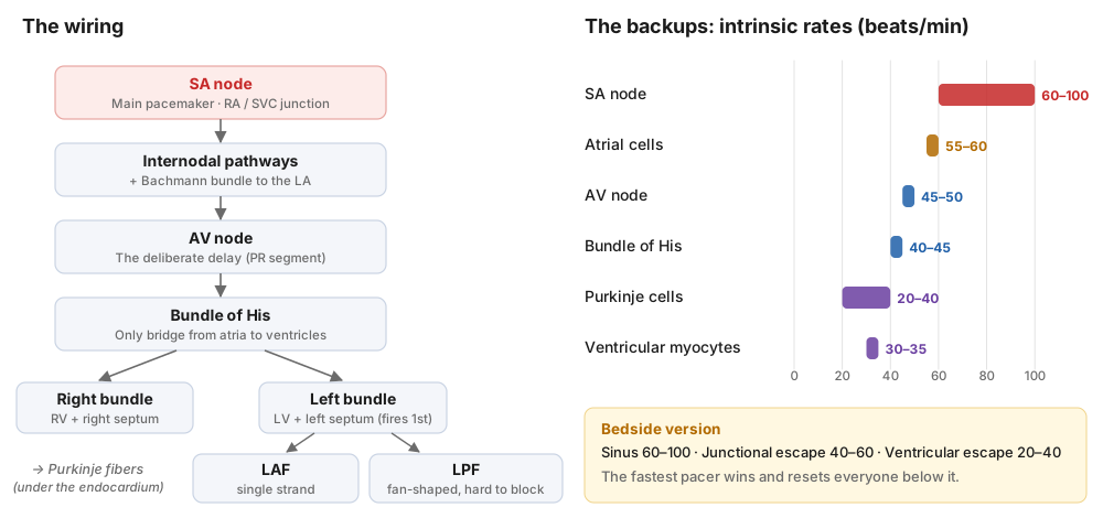
- SA node: the main pacemaker, at the junction of the right atrium and SVC. Fires at 60–100 (average ~70). Blood supply is the RCA in ~60% of people.
- Internodal pathways + Bachmann bundle: carry the impulse across the atria to the AV node and over to the left atrium.
- AV node: slows conduction on purpose so the atria can top off the ventricles. Usually supplied by the RCA, which is why inferior MIs cause AV block.
- Bundle of His: the only electrical bridge between atria and ventricles.
- Right bundle: RV and the right side of the septum.
- Left bundle: LV and the left side of the septum. Its septal fibers fire first (that's the small "septal q" in the lateral leads).
- Left anterior fascicle (LAF): one thin strand, so it blocks easily.
- Left posterior fascicle (LPF): fan-shaped and widely spread, so it is hard to block.
- Purkinje fibers: sit just under the endocardium and start ventricular depolarization.
Core Principle: Every cell in the system can pace, but each one down the line is slower. If the SA node fails, the next fastest site takes over. Bedside version: sinus 60–100, junctional escape 40–60, ventricular escape 20–40.
2. The Spark: Electrophysiology
A resting myocyte sits at about −70 to −90 mV. The Na⁺/K⁺ ATPase pump keeps it that way by moving 3 Na⁺ out for every 2 K⁺ in. Sodium is high outside the cell; potassium is high inside.
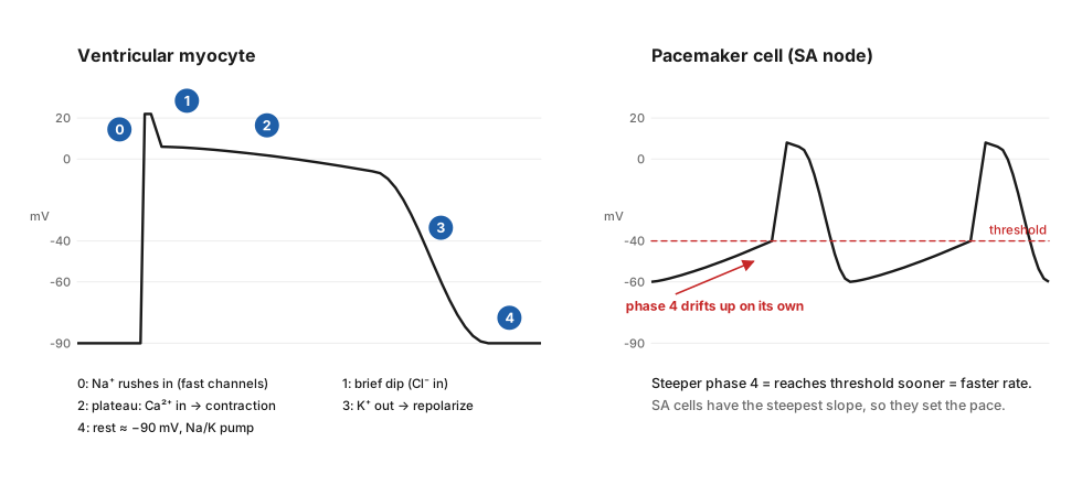
Phase | What happens | ECG link |
0 | Fast Na⁺ channels open → rapid upstroke | QRS |
1 | Brief dip as the fast channels close | J point |
2 | Plateau: Ca²⁺ (and slow Na⁺) flow in and keep the cell depolarized | ST segment |
3 | K⁺ flows out → rapid repolarization | T wave |
4 | Rest; pump restores the gradients | TP baseline |
Calcium is the key to contraction. It unlocks troponin/tropomyosin so actin and myosin can slide past each other.
Why phase 4 matters: pacemaker cells don't sit still at rest; they slowly leak toward threshold. The steeper the slope, the faster the rate. SA cells have the steepest slope, so they win.
Why EM cares: the same ions drive the tracing. High K⁺ → peaked T waves. Low Ca²⁺ or Mg²⁺ → long QT.
3. Vectors, Leads and Territories
Each depolarizing cell creates a small force with a size and a direction (a vector). Add them all together and you get the heart's electrical axis (more detail in  ECG Topic - Electrical Axis).
Each lead is a camera looking at that vector from one angle.
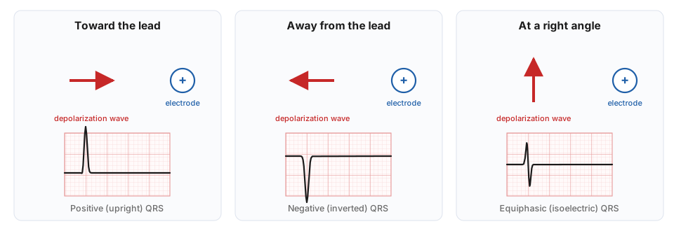
Two lead systems:
- Hexaxial (limb leads: I, II, III, aVR, aVL, aVF): look at the heart in the frontal plane (up/down, left/right).
- Precordial (V1–V6): look in the horizontal plane (front/back, left/right).
Together they give a 3-D view. That's what lets you localize ischemia:
Territory | Leads | Usual artery |
Inferior | II, III, aVF | RCA (most), LCx |
Septal | V1, V2 | LAD (septal branches) |
Anterior | V3, V4 | LAD |
Lateral | I, aVL, V5, V6 | LCx, LAD diagonals |
Combinations are common: inferolateral = inferior + lateral leads.
4. The Paper
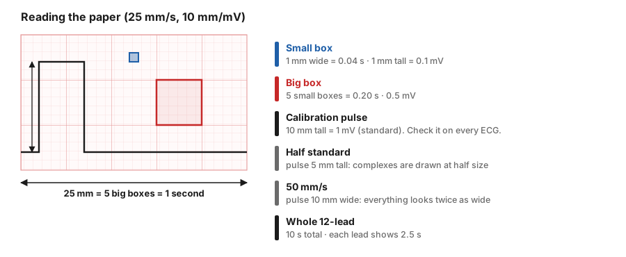
- Small box: 1 mm = 0.04 s wide, 0.1 mV tall.
- Big box: 5 small boxes = 0.20 s.
- 5 big boxes = 1 second.
- Calibration pulse: 10 mm tall = 1 mV. A 5 mm pulse means half standard (everything looks half size). A pulse 10 mm wide means the paper ran at 50 mm/s (everything looks twice as wide).
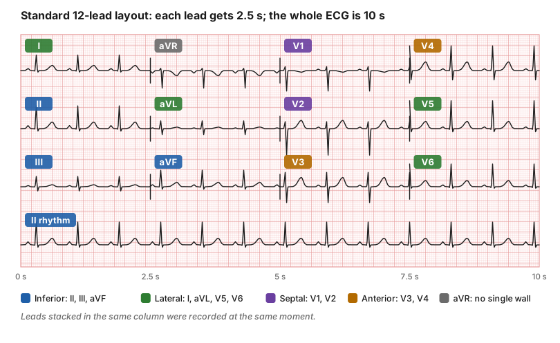
Why the timing layout matters: the leads in one column happened at the same time. If the start of a QRS is unclear in one lead, look at another lead in the same column. A wave that looks odd in one lead can be checked against its neighbors.
Tools (Garcia Ch. 5): calipers are your best friend. Use them to compare R-R and P-P intervals, check whether an interval is fixed or changing, and "march out" P waves hidden in QRS complexes or T waves.
5. The Basic Beat
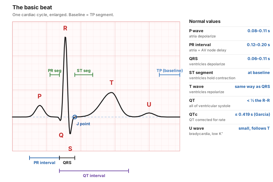
Component | What it means | Normal |
P wave | Atrial depolarization | 0.08–0.11 s; upright in I and II |
PR interval | Start of P → start of QRS (atria + AV node) | 0.12–0.20 s. ≤ 0.11 = short; ≥ 0.21 = 1st-degree block |
QRS | Ventricular depolarization | 0.06–0.11 s |
Q wave | First downward deflection of the QRS | Pathologic if ≥ 0.03 s wide or ≥ ⅓ the R-wave height |
J point | Where the QRS ends and the ST begins | Where you measure ST deviation |
ST segment | Electrically quiet; ventricles stay contracted | At baseline. Up to 1 mm in limb leads and up to 3 mm in V1–V3 can be normal (early repolarization, LVH) |
T wave | Ventricular repolarization | Same direction as QRS; asymmetric (slow up, fast down) |
QT interval | All of ventricular systole | Less than half the preceding R-R |
U wave | Small wave after T | Seen with bradycardia and low K⁺ |
Why is the T wave upright when repolarization is a "negative" wave? Depolarization runs endocardium → epicardium. Repolarization runs the other way, epicardium → endocardium. A negative wave moving away from a lead looks the same as a positive wave moving toward it. So the T follows the QRS.
Symmetric T waves can be normal, but usually mean pathology (ischemia, hyperkalemia).
QTc: the QT shortens as the rate rises, so correct it for rate. Garcia uses QTc = QT + 1.75 × (HR − 60), with > 0.419 s (419 ms) as prolonged. Most machines use Bazett (QT ÷ √RR), which overcorrects at fast rates. Many references use > 450 ms (men) and > 460 ms (women) as prolonged, and > 500 ms as high risk for torsades.
⚠️ Don't Miss: ST elevation that doesn't meet STEMI criteria isn't automatically benign. Get an old ECG and repeat the tracing.
Pearl: ECG machines often include the U wave in the QT and overcall a long QT. Measure it yourself.
6. The Rate
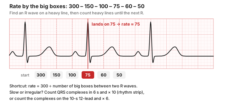
Method | Use when | How |
300 – 150 – 100 – 75 – 60 – 50 | Regular rhythm | R wave on a heavy line; name each heavy line after it until the next R |
300 ÷ big boxes | Regular rhythm | Count big boxes between two R waves |
6-second strip × 10 | Slow or irregular | Count QRS complexes in 6 s, multiply by 10 |
12-lead × 6 | Any rhythm on a 12-lead | Count QRS complexes across the full 10 s, multiply by 6 |
Use the right math for the right strip. A rhythm strip has 3-second hash marks. A 12-lead is 10 seconds long, and each lead shows only 2.5 seconds.
7. Rhythms
The 10 Questions
Garcia's system for any rhythm. Ask them in order, every time.
# | Question | Why it matters |
1 | Fast or slow? | > 100 tachy, < 60 brady. Many rhythms live in set rate ranges. |
2 | Regular or irregular? If irregular, regularly or irregularly irregular? | Only three rhythms are irregularly irregular (below). |
3 | Any P waves? | P waves = an atrial/supraventricular component. |
4 | Are all the P waves the same? | Different P waves = a different pacer (or a T wave hiding in the P). ≥ 3 shapes = WAP or MAT. |
5 | A P for every QRS? | More Ps than QRSs → think AV block. |
6 | Is the PR constant? | Changing PR → Wenckebach, WAP/MAT, or dissociation. |
7 | Are P and QRS related? | No relation = AV dissociation or complete heart block. |
8 | Narrow or wide QRS? | Narrow = used the normal His-Purkinje system. Wide = ventricular origin, aberrancy or bundle branch block. |
9 | Grouped beats? | Grouping → Wenckebach, bigeminy or trigeminy. |
10 | Dropped beats? | AV block, SA block, or sinus arrest. |
Memorize this: only three rhythms are irregularly irregular: atrial fibrillation, wandering atrial pacemaker and multifocal atrial tachycardia. No P waves → AFib. ≥ 3 different P waves → WAP (< 100) or MAT (> 100).
Sinus Rhythms
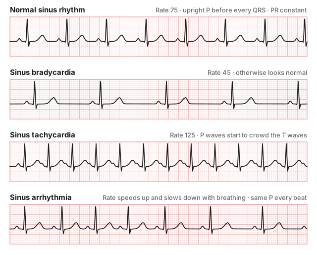
Rhythm | Key features |
Normal sinus rhythm | 60–100, regular, upright P before every QRS |
Sinus bradycardia | < 60, otherwise normal |
Sinus tachycardia | > 100, otherwise normal. Look for the cause (pain, fever, bleeding, PE, sepsis). |
Sinus arrhythmia | Rate rises and falls with breathing. A normal variant, especially in young people. |
Sinus pause / arrest | The SA node stops; the next beat comes at no set time. |
SA block | A whole P-QRS-T drops, but the next beat lands exactly where the rhythm predicts (pause = a multiple of the P-P interval). |
Atrial Rhythms
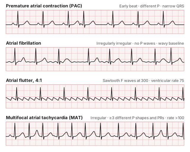
Rhythm | Key features |
PAC | Early beat with a different-looking P; narrow QRS (unless aberrant) |
Ectopic atrial tachycardia | Regular, > 100, all P waves the same but not sinus-shaped |
Wandering atrial pacemaker | ≥ 3 P shapes and PRs, rate < 100 |
MAT | ≥ 3 P shapes and PRs, rate > 100. Think COPD and lung disease. |
Atrial flutter | Sawtooth F waves at ~250–350/min (usually ~300); often 2:1 block → ventricular rate ~150 |
Atrial fibrillation | Irregularly irregular, no P waves, wavy or flat baseline |
Pearl: a regular, narrow-complex tachycardia at ~150 is flutter with 2:1 block until proven otherwise. Hunt for the second F wave hiding in the T wave or QRS.
Junctional Rhythms
The AV junction takes over. QRS is narrow. The P wave is absent, or inverted with a short PR (before the QRS), or appears just after the QRS.
Rhythm | Rate |
PJC | Single early junctional beat |
Junctional escape beat | Late beat that fills a pause |
Junctional rhythm | 40–60 |
Accelerated junctional | 60–100 |
Junctional tachycardia | > 100 |
Ventricular Rhythms
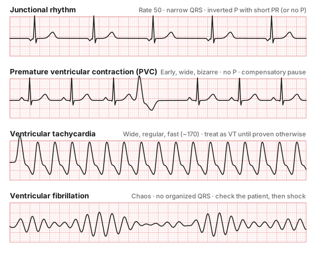
Rhythm | Key features |
PVC | Early, wide, bizarre; no P; T wave points opposite the QRS; usually a full compensatory pause |
Ventricular escape beat | Late wide beat that rescues a pause |
Idioventricular rhythm | Wide, regular, 20–40 |
Accelerated idioventricular (AIVR) | Wide, regular, 40–100. Classic after reperfusion. |
Ventricular tachycardia | Wide, > 100, usually regular |
Torsades de pointes | Polymorphic VT that twists around the baseline; associated with long QT |
Ventricular flutter | Sine wave at ~300. Think WPW with 1:1 flutter conduction. |
Ventricular fibrillation | Chaotic, no organized QRS |
VT clues:
- AV dissociation (P waves marching through)
- Fusion and capture beats (hallmarks of VT)
- QRS ≥ 0.16 s
- All precordial leads negative (negative concordance)
- Brugada sign: R to the bottom of the S ≥ 0.10 s
- Josephson sign: notching near the bottom of the S wave
⚠️ Wide-complex tachycardia = VT until proven otherwise. Calling it SVT with aberrancy is a common and dangerous mistake.
⚠️ Never suppress an escape rhythm. Idioventricular rhythm or AIVR may be the patient's last pacemaker. Treat it with antiarrhythmics and you get asystole.
Pearl: "VFib" in an awake patient talking to you is a loose lead.
AV Blocks
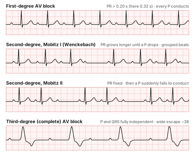
Block | PR interval | Dropped beats? |
First-degree | > 0.20 s, constant | No; every P conducts |
Mobitz I (Wenckebach) | Gets longer each beat, then a P drops | Yes, in groups. Usually at the AV node, usually benign. |
Mobitz II | Constant, then a P suddenly drops | Yes. Below the AV node; can progress to complete block. |
Third-degree (complete) | No relationship | Ps and QRSs each run at their own rate; atrial rate > ventricular rate |
⚠️ 2:1 block? You can't tell Mobitz I from Mobitz II when every other P drops. Call it "2:1 second-degree block" and treat it like Mobitz II.
Semantics: if the P waves and QRSs are dissociated but there are as many or more QRSs as Ps, it's AV dissociation, not complete heart block. And AV blocks are not bundle branch blocks; those are a different problem.
High-Yield Pearls
- Fastest pacer wins. Escape rhythms are backups, so look for why the higher pacer failed.
- Check the calibration pulse first. Half standard or 50 mm/s will fool every measurement after it.
- Same column = same moment. Use neighboring leads to find the true start and end of a wave.
- Intervals include waves; segments don't. PR interval includes the P; PR segment doesn't.
- QRS > 0.11 s? The impulse took a detour: bundle branch block, ventricular origin or aberrancy.
- T waves should follow the QRS and be asymmetric. Symmetric or opposite → look for a reason.
- Three irregularly irregular rhythms: AFib, WAP, MAT.
- Regular narrow tachycardia at 150 → hunt for flutter waves.
- Wide and fast = VT until proven otherwise.
- 2:1 AV block → treat like Mobitz II.
- Don't treat escape rhythms with antiarrhythmics.
Quick Algorithm
- Calibration: 25 mm/s and 10 mm/mV?
- Rate: 300 sequence (regular) or count × 10 / × 6 (irregular).
- Regular? If not, regularly or irregularly irregular?
- P waves: present? all the same? one for every QRS?
- PR: normal (0.12–0.20), short, long, changing?
- QRS: narrow (≤ 0.11) or wide?
- P–QRS relationship: linked, grouped, dropped or dissociated?
- Name the rhythm, then move on to the rest of the 12-lead (intervals, axis, ST/T).
Practice - Rate and Rhythm
Try each one before opening the answer.
Part A - Rate
# | Finding |
A1 | Regular rhythm, 3 big boxes between R waves |
A2 | Regular rhythm, 5 big boxes between R waves |
A3 | Regular rhythm, 2 big boxes between R waves |
A4 | Irregular rhythm, 4 QRS complexes on a 6-second strip |
A5 | Irregular rhythm, 14 QRS complexes on a full 12-lead |
A6 | 3.5 beats in a 6-second strip |
Part B - Name That Rhythm
Use the 10 questions. Each strip is lead II at standard settings.
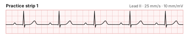
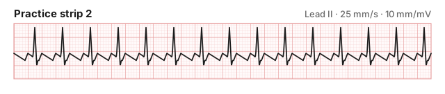
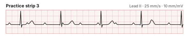
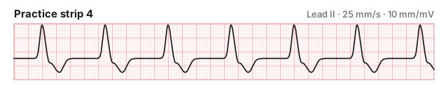
Part C - Concept Check
- What is the only electrical connection between the atria and ventricles?
- Which fascicle is hardest to block, and why?
- What ion's influx creates the phase 2 plateau and triggers contraction?
- A wave moves away from a lead. What does the lead record?
- The calibration pulse is 5 mm tall. What does that mean?
- Name the three irregularly irregular rhythms.
- Every other P wave is dropped. Mobitz I or II?
Sources
- Garcia TB. 12-Lead ECG: The Art of Interpretation. 2nd ed. Burlington, MA: Jones & Bartlett Learning; 2015. Chapters 1–8.
This post is for education and not medical advice.