

Last Updated November 2026
Introduction
The left bundle splits into two fascicles: the left anterior fascicle and the left posterior fascicle. Block one of them and you get a fascicular block. The QRS stays narrow, but the axis swings hard to one side. In the ED you need to answer three questions: Is the axis abnormal? Is it a fascicular block? Is there also an RBBB?
The Anatomy in One Picture
- Left anterior fascicle: a thin, tight cord. It supplies the anterior and lateral walls of the LV. Easy to block, so LAFB is common.
- Left posterior fascicle: loose fibers that fan out over the inferior and posterior walls. You need a big lesion to block it. Hard to block, so LPFB is rare.
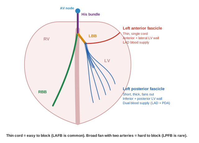
Core Principle: The blocked part of the LV fires last, and nothing cancels it. That late force pulls the axis toward the blocked wall. Block the anterior (upper) fascicle → axis swings up and left. Block the posterior (lower) fascicle → axis swings down and right.
Left Anterior Fascicular Block (LAFB)
What Happens
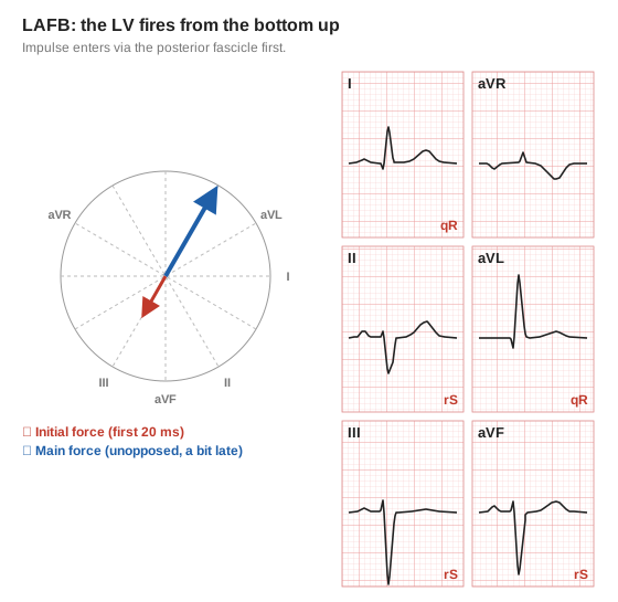
The impulse enters the LV through the posterior fascicle, so the LV fires from the bottom up.
- First: a small force points down and right → small r in II, III, aVF and a small q in I, aVL.
- Then: a big, late force points up and left → tall R in I, aVL and a deep S in II, III, aVF.
LAFB Criteria
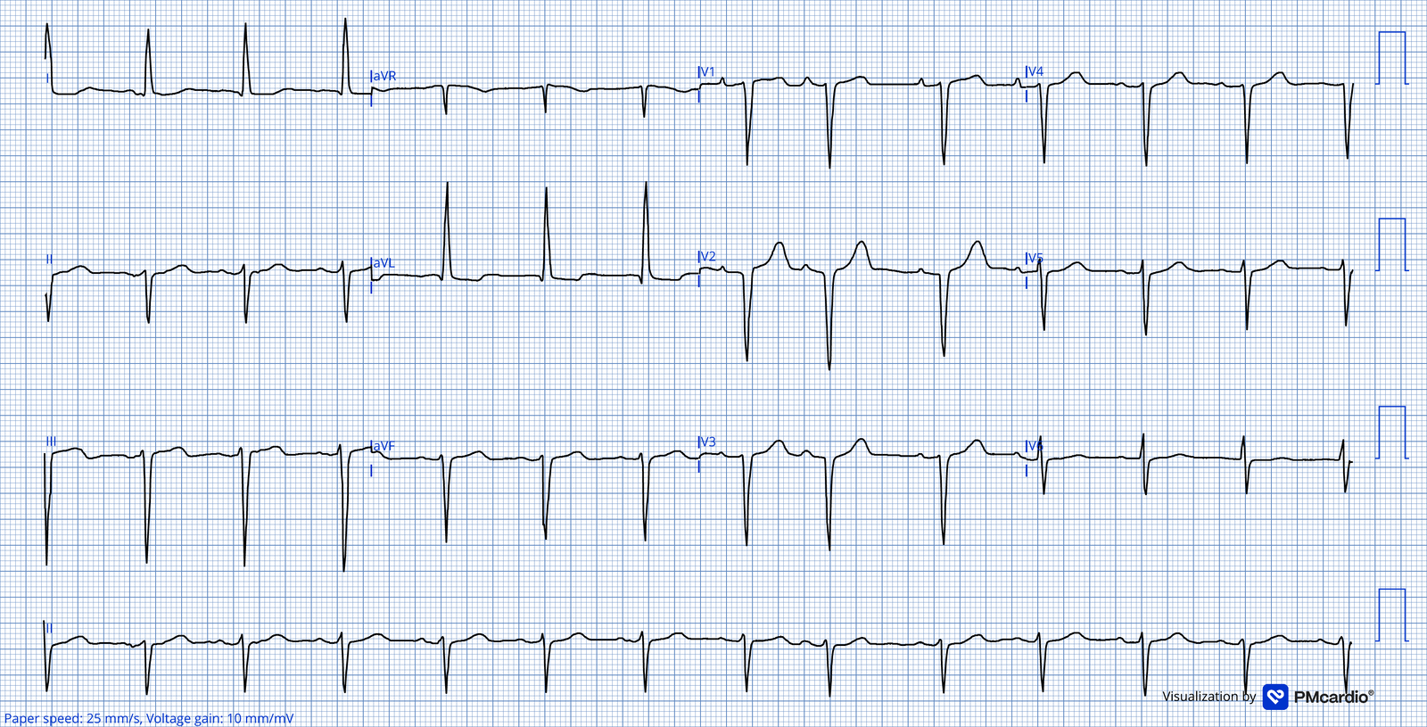
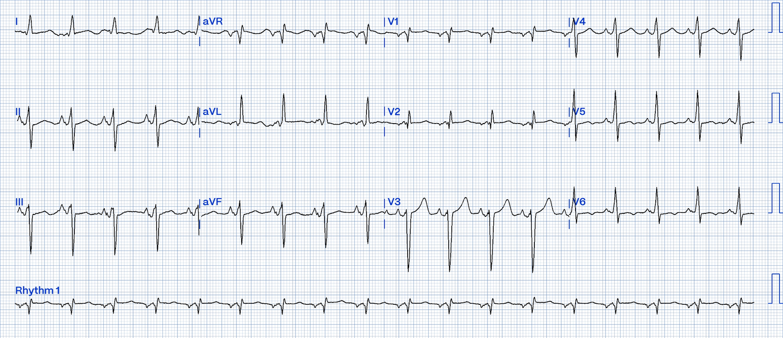
- Left axis deviation, specfically −30° to −90°
- qR (or a tall R) in lead I
- rS in lead III (and usually II and aVF)
The 2-Second Left Axis Check
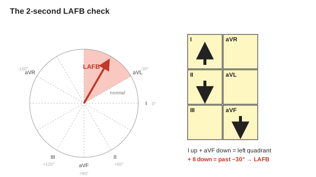
- Lead I up
- aVF down
- Lead II down
That combination only fits an axis between −30° and −90°. With a narrow QRS, that's LAFB.
Garcia's tip: The small r in lead III can be tiny. Look closely. It doesn't matter how big a wave is, just that it's there.
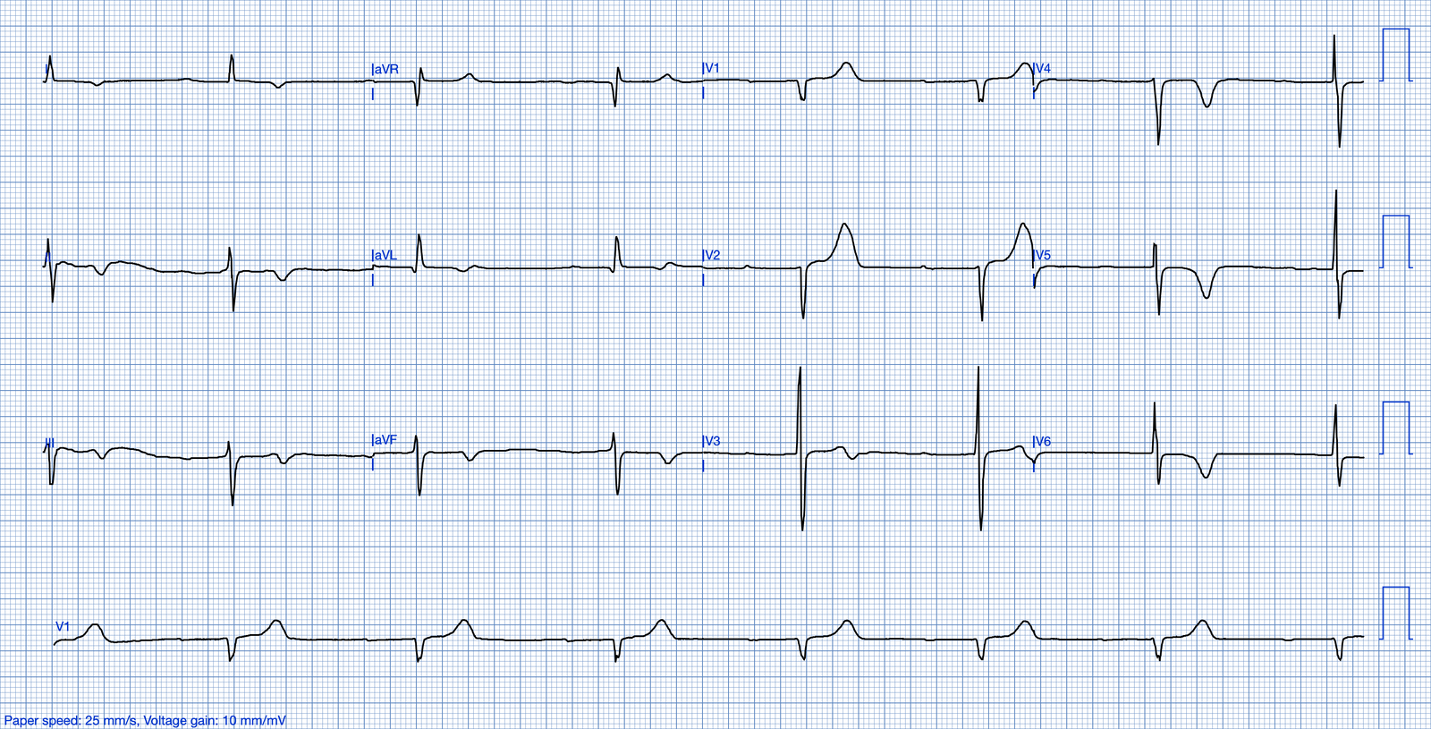
Examples of LAFB
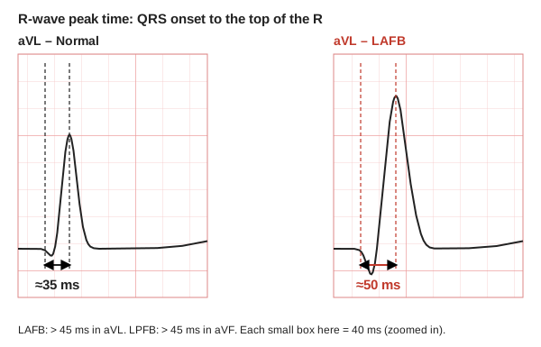
Bonus: R-Wave Peak Time (LITFL)
Measure from the start of the QRS to the top of the R in aVL. In LAFB it's more than 45 ms, because that late force arrives late. Useful when the axis is borderline.
LAFB vs Inferior MI
Both give you a left axis. Look at the start of the QRS in II, III and aVF:
- LAFB: starts with a small r, then a deep S (rS).
- Inferior MI: starts with a Q (Qr or QS).
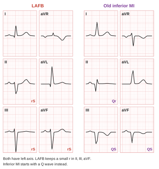
Some people say you can't call LAFB when there are inferior Q waves. An axis between −30° and −90° is abnormal no matter what, and a Q wave can hide the small r. If you miss a fascicular block in an MI, the patient can go into complete heart block. If you overcall it, no one is harmed. Don't be too strict.
Don't Be Fooled
- In LAFB, the voltage in lead aVL may meet voltage criteria for LVH (R wave height > 11 mm).
- Tall R in aVL ≠ LVH. LAFB points the main force straight at aVL, so the R can get tall (over 11 mm). With no strain pattern, be careful calling LVH.
- LAFB is common and often stable. A new LAFB in a patient with chest pain is different: think ischemia.
Left Posterior Fascicular Block (LPFB)
What Happens
The impulse enters the LV through the anterior fascicle, so the LV fires from the top down. It's the mirror image of LAFB.
- First: a small force points up and left → small r in I, aVL and a small q in II, III, aVF.
- Then: a big, late force points down and right → deep S in I, aVL and a tall R in II, III, aVF.
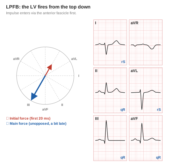
Criteria (Garcia)
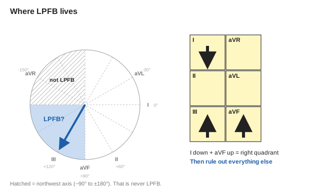
- Axis +90° to +180° (the right quadrant, not the extreme right/northwest)
- S in lead I and q in lead III
- No RAE and no RVH
Examples of LPFB
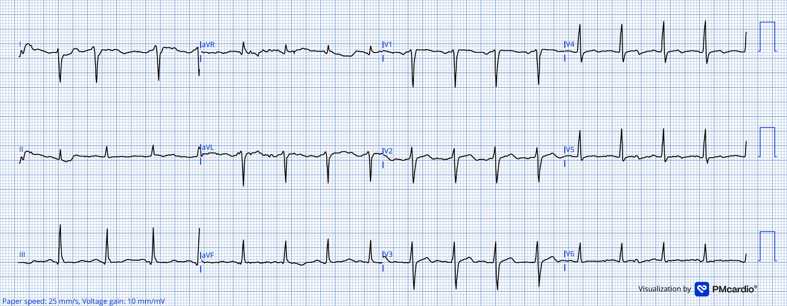
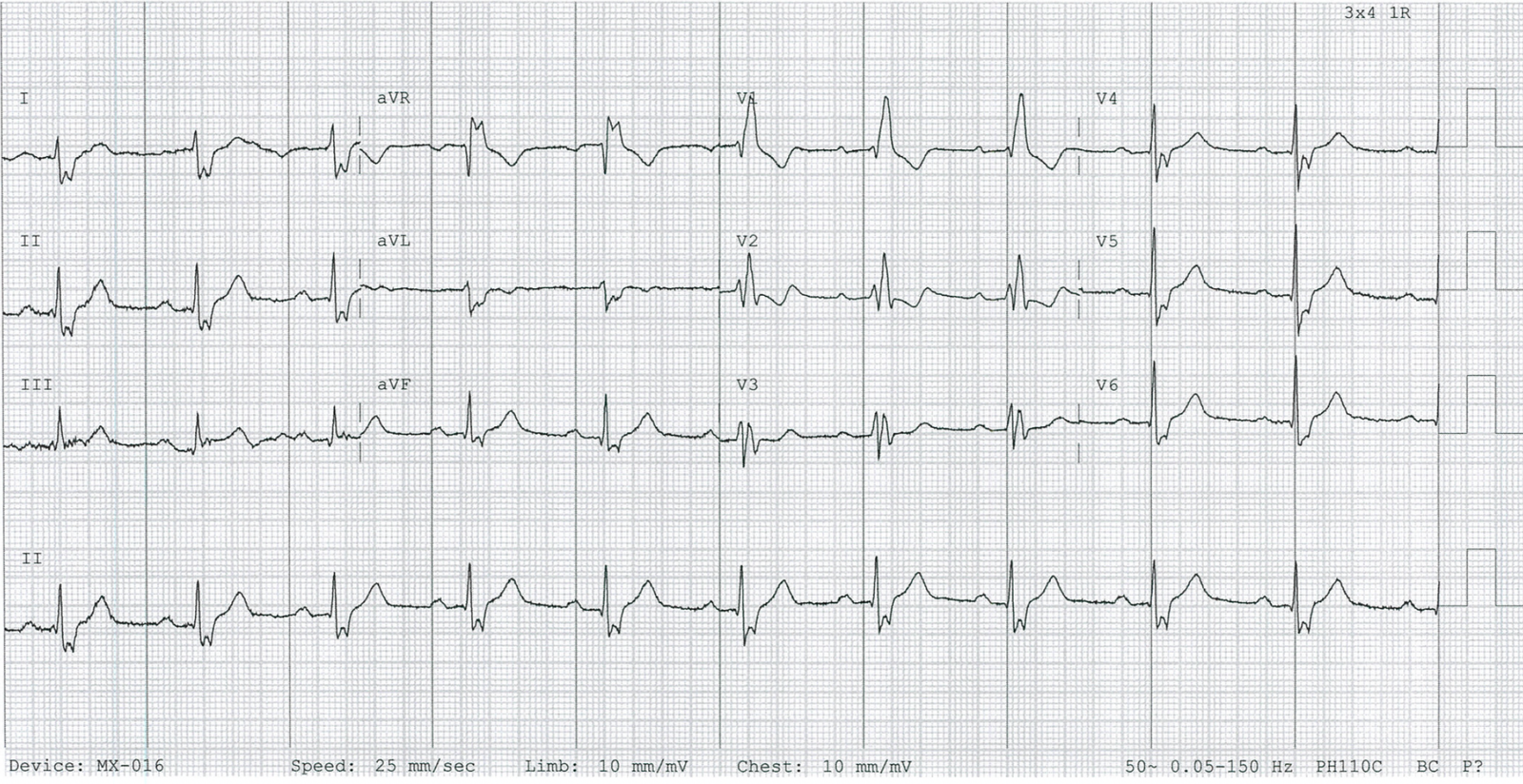
It's a Diagnosis of Exclusion
No finding proves LPFB. You get there by ruling out everything else that causes a right axis. You could read thousands of ECGs and be lucky to find one.
Rule out | Look for |
RVH (most common cause of RAD) | Tall R in V1, TWI in V1–V3 |
Right atrial enlargement | P wave ≥ 2.5 mm tall in II (P pulmonale) |
Pulmonary embolism | Tachycardia, S1Q3T3. Garcia: the ECG alone often can't tell PE from LPFB. You need the history. |
Lateral MI | Q waves in I, aVL |
Others (LITFL) | TCA overdose, normal variant in young/thin patients, lead reversal |
Every time you see a right axis, ask yourself: "Is this LPFB?" If you don't think about it, you'll miss it. Then rule out RVH, RAE, PE and lateral MI before you call it. S1Q3T3 is not pathognomonic for PE. It is suggestive. It can also come from LPFB or RVH
Side by Side
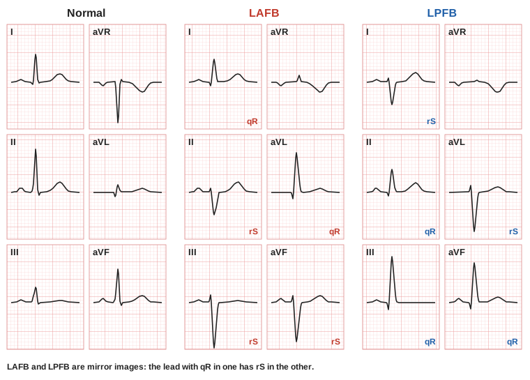
LAFB | LPFB | |
Axis | −30° to −90°
(up in I and down in II & aVF) | +90° to +180°
(down in I and up in III & avF) |
Lead I | qR | rS |
Lead III | rS | qR |
How common | Common | Rare |
How you call it | Rule it in | Rule everything else out |
Bifascicular Block
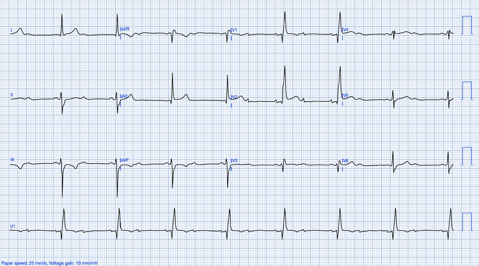
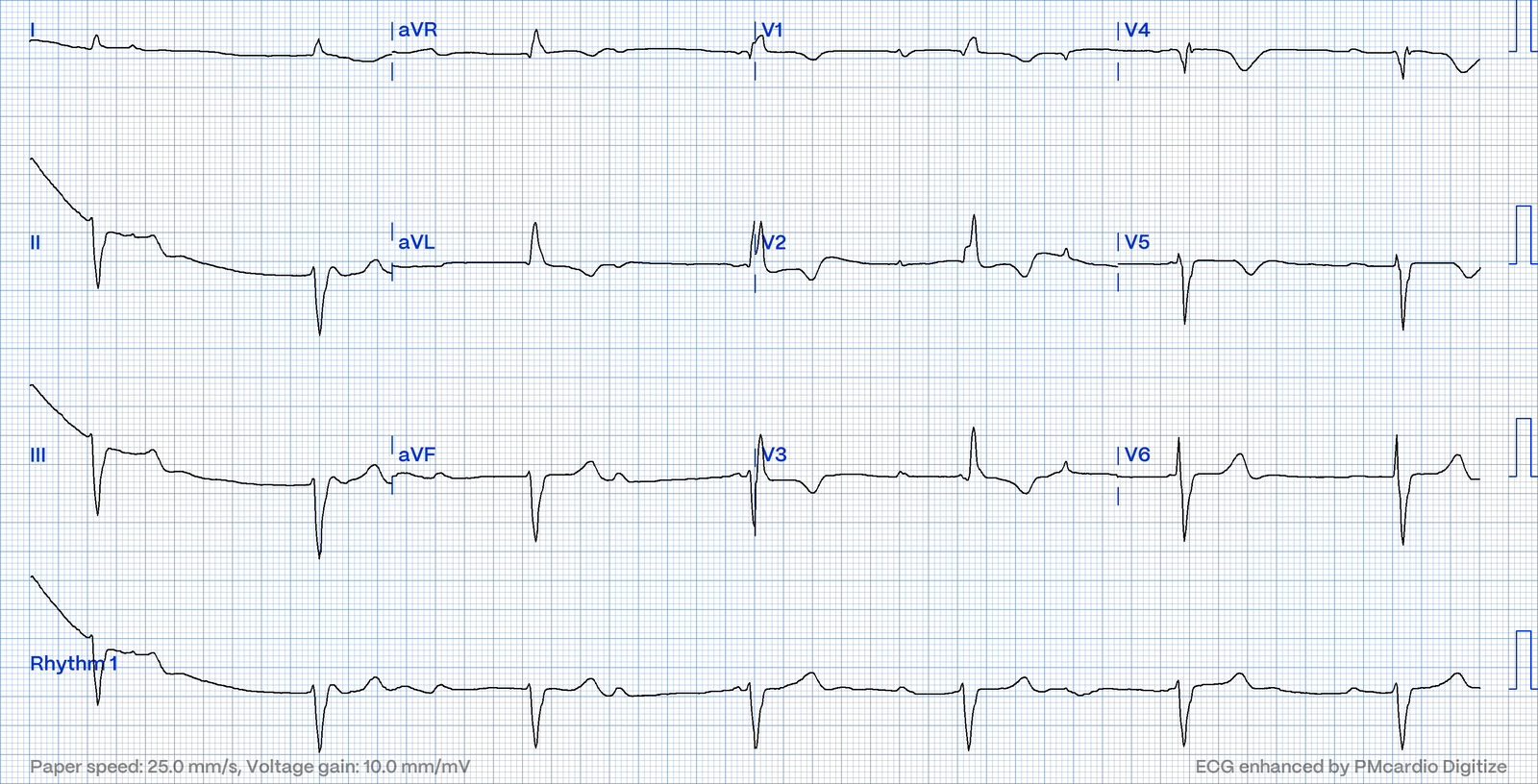
Three fascicles carry the impulse: the right bundle, the left anterior fascicle and the left posterior fascicle. A bifascicular block is RBBB + LAFB or RBBB + LPFB. Now one fascicle carries every beat.
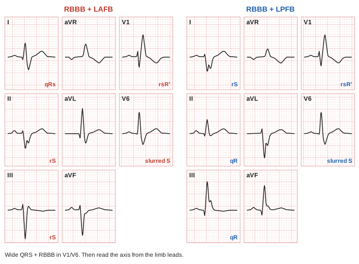
How to Read It (Garcia's Steps)
- QRS ≥ 120 ms?
- Slurred S in I and V6? RSR′ in V1? → RBBB.
- Now do the axis:
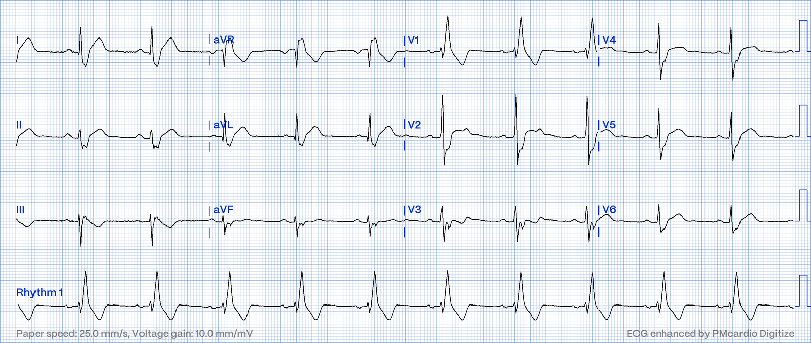
- I up, aVF down, II down → RBBB + LAFB
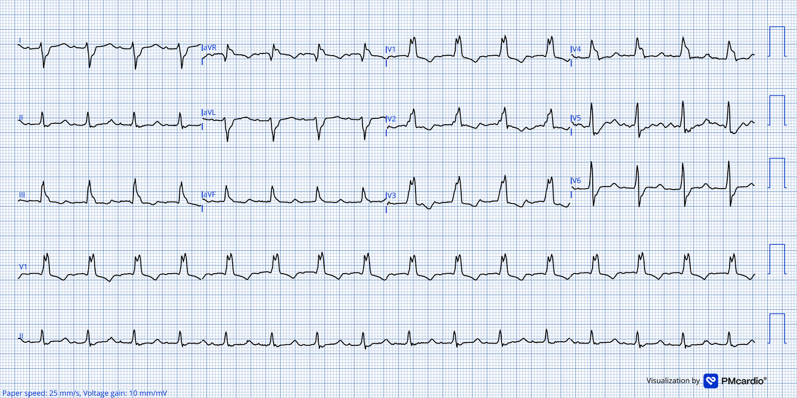
- I down, aVF up, q in III (and no RVH/RAE) → RBBB + LPFB
A positive QRS in lead I with a slurred S means one of two things: RBBB with a normal axis, or RBBB + LAFB. Check lead II.
Which One Should Worry You?
RBBB + LAFB | RBBB + LPFB | |
How common | Common | Less common (but more common than LPFB alone) |
Stable? | Yes, chronically | No. Only the thin anterior fascicle is left |
During an acute MI | Can progress to complete heart block | High risk of complete heart block |
⚠️ Don't Miss: Bifascicular block in an acute MI can turn into complete heart block. Garcia treats this as an indication for pacing. Get pads on and the pacer ready before the patient crashes.
Examples
Bifasicular Block - RBBB+LAFB
Bifasicular Block - RBBB+LPFB
Bifascicular block (RBBB+LAFB) w/ 2nd degree degree av block
Bifascicular block (RBBB+LAFB) w/ 3rd degree degree av block
High-Yield Pearls
- Narrow QRS + big axis shift = think fascicular block.
- I up, aVF down, II down = LAFB. Takes 2 seconds.
- Inferior Q waves + left axis? Think inferior MI, but don't be too strict about calling LAFB too.
- Right axis? Ask "Is this LPFB?" Then rule out RVH, RAE, PE and lateral MI.
- Northwest axis is never LPFB.
- Wide QRS? Find the RBBB first, then check the axis for a bifascicular block.
- RBBB + LPFB is the unstable one. Any bifascicular block in an acute MI → be ready to pace.
Quick Algorithm
- Look at Lead I and aVF.
- I up, aVF down → check Lead II.
- II down → LAFB (if the inferior leads start with a small r).
- II up → normal (0° to −30°).
- I down, aVF up → right axis → "Is this LPFB?" Rule out RVH, RAE, PE, lateral MI.
- Both down → northwest axis. Not a fascicular block.
- QRS wide? Look for RBBB → bifascicular block?
Practice - Name That Block
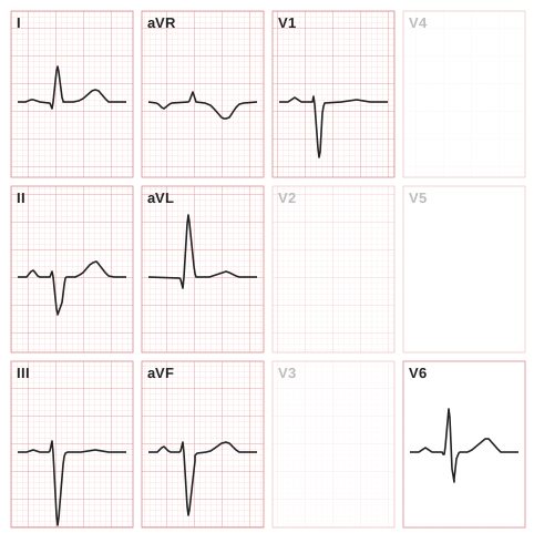
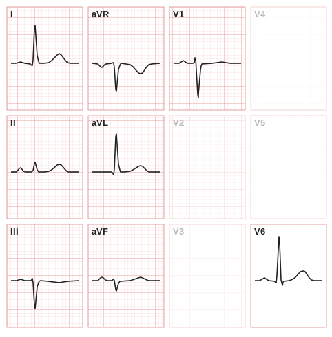
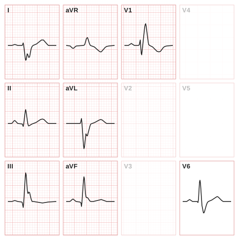
These are drawn single beats in a 12-lead layout. V2–V5 are left blank because you don't need them for these cases. For each one: quadrant → QRS shapes → V1/V6 → name it.
Exercise 1
Exercise 2
Exercise 3
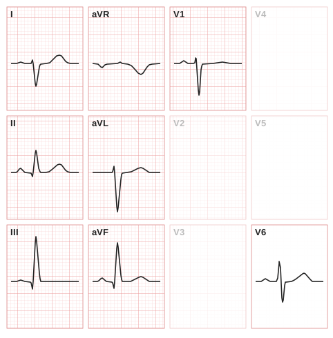
Exercise 4
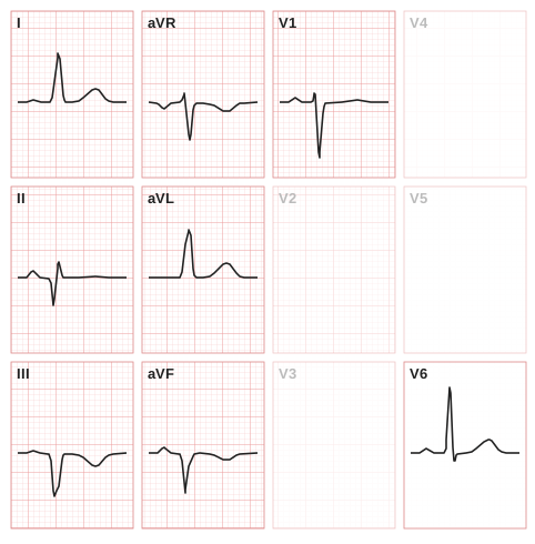
Exercise 5
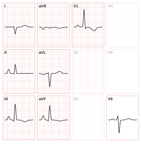
Exercise 6
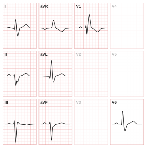
Exercise 7
Sources
- Garcia TB. 12-Lead ECG: The Art of Interpretation. 2nd ed. Burlington, MA: Jones & Bartlett Learning; 2015. Chapter 13, Bundle Branch Blocks and Hemiblocks.
- Burns E, Buttner R. Left Anterior Fascicular Block (LAFB). Life in the Fast Lane ECG Library. https://litfl.com/left-anterior-fascicular-block-lafb-ecg-library/
- Burns E, Buttner R. Left Posterior Fascicular Block (LPFB). Life in the Fast Lane ECG Library. https://litfl.com/left-posterior-fascicular-block-lpfb-ecg-library/
This post is for education and not medical advice.