

Last Updated November 2026
Introduction
Paced ECGs intimidate most non-cardiologists. Pacemakers have advanced names, dozens of programmable features, and rhythm strips that look broken when they are actually working perfectly, a lot of info which might not be relevant knowledge when working in the ER.
A pacemaker has 2 major roles - it senses and it paces. Every paced strip, normal or abnormal, can be explained by asking how well the device is doing those two jobs. This post is a non-academic approach for the non-cardiologist: learn the language (the mode), recognize when sensing or pacing goes wrong, and recognize the features that look abnormal but may be completely normal.
Core Principle: The pacemaker looks for a native impulse. Present → inhibit (stay quiet). Absent → pace. Almost every abnormal paced strip is a sensing problem, a capture problem, or a programmed feature doing exactly what it was designed to do.
Paced QRS Morphology
Where the ventricular lead sits determines what the paced QRS looks like. Paced beats are wide because the impulse travels muscle-to-muscle instead of through the conduction system.
Lead location | Morphology | Why |
RV pacing (most common) | LBBB-like • wide, negative in V1 | Depolarization starts in the RV and spreads right → left |
LV pacing | RBBB-like • wide, positive in V1 | Depolarization starts in the LV and spreads left → right |
Biventricular (CRT) | RBBB-ish, often narrower than RV pacing; frequently negative in lead I | Both ventricles are stimulated nearly simultaneously |
RV paced rhythm with LBBB morphology

Source: LITFL
OMI Pearl: RV paced rhythm hides ischemia the same way LBBB does. Use the Smith-Modified Sgarbossa criteria (concordant STE ≥1 mm, concordant STD ≥1 mm in V1-V3, or excessively discordant STE with ST/S ratio ≥25%) to look for occlusion MI in a paced rhythm.
BiV paced rhythm with RBBB morphology

super:{{ eager }}
Source: ECG Stampede
Pacemaker Modes (NBG Code)
The mode is the pacemaker's language. It is written as three letters: chamber paced → chamber sensed → response to sensing.
Position | I - Chamber Paced | II - Chamber Sensed | III - Response to Sense |
Options | Atrium | Atrium | Inhibit |
Ventricle | Ventricle | Trigger | |
Dual (A + V) | Dual (A + V) | Dual (I + T) | |
- | O (none) | O (none) |
When you look at a paced strip, fill in the three blanks:
- Chamber paced - where are the spikes? Atrium, ventricle, or both?
- Chamber sensed - do native beats in that chamber reset the device?
- Sense response - does a sensed beat inhibit pacing, trigger pacing, or both?
Single Chamber Modes
VVI - Ventricular Demand Pacing
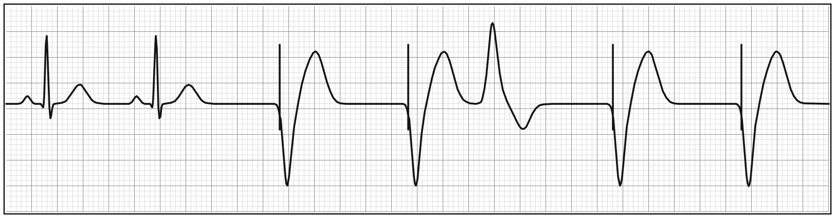
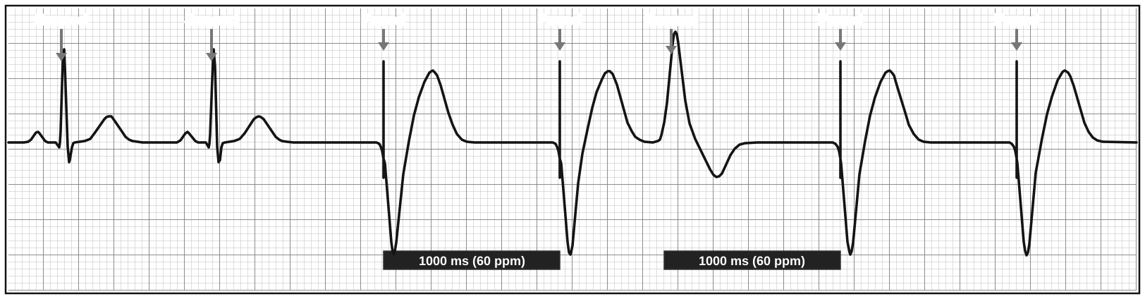
- Paced: V · Sensed: V · Response: Inhibit
- Two native beats are sensed (no spikes). When the native rhythm stops, the device paces at the base rate (1000 ms = 60 ppm)
- The PVC is sensed - the next paced beat comes exactly one pacing interval after it
- VVI ignores the atrium completely, so it provides no AV synchrony
VOO - Asynchronous Ventricular Pacing
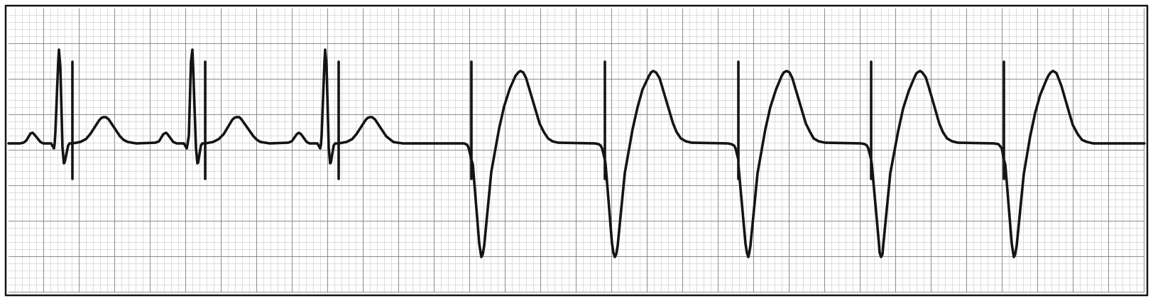
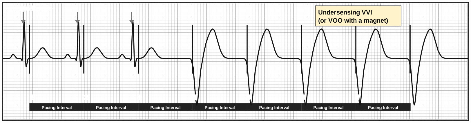
- Paced: V · Sensed: none · Response: none
- Spikes march at a fixed pacing interval no matter what the heart does - spikes 1-3 land right after native QRS complexes the device never saw
- This is how a pacemaker behaves with a magnet on it, during surgery/MRI programming, or when it has reverted for noise
- A VOO strip and an undersensing VVI look identical. If no one put a magnet on or reprogrammed it, think undersensing
R on T Phenomenon
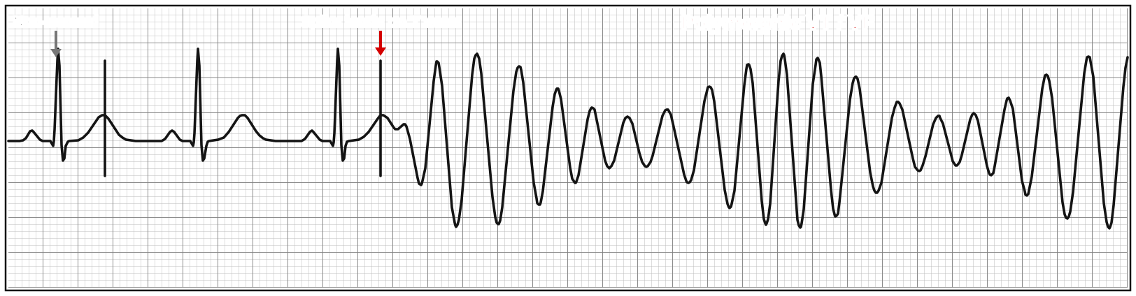
- When the device doesn't sense native beats, a spike can land on the T wave (the vulnerable period)
- Rarely, this triggers polymorphic VT / VF
AAI - Atrial Demand Pacing
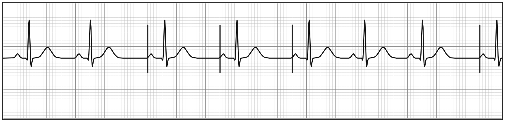
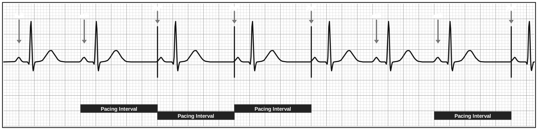
- Paced: A · Sensed: A · Response: Inhibit
- Native P waves (AS) inhibit the device; when the sinus node pauses, the device paces the atrium (AP)
- Atrial spike → P wave → narrow, native QRS. The patient's own AV node conducts
- Used for isolated sinus node dysfunction with a healthy AV node
AOO - Asynchronous Atrial Pacing
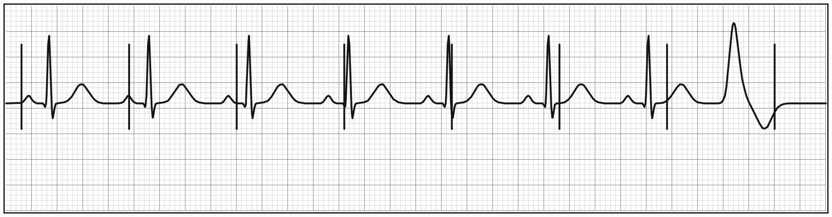
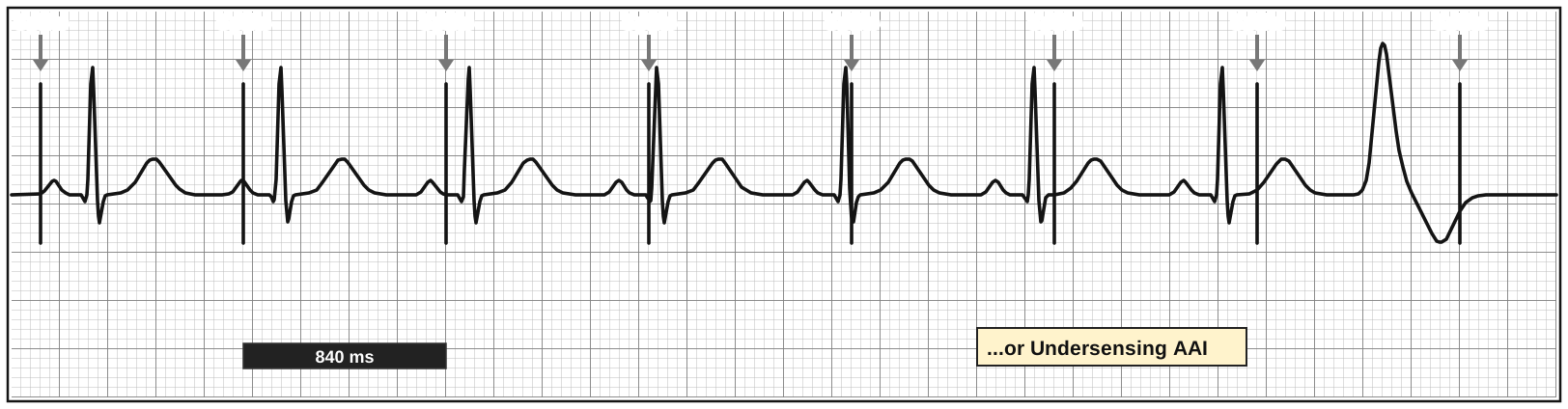
- Paced: A · Sensed: none · Response: none
- Atrial spikes march every 840 ms regardless of the native P waves and QRS complexes
- Like VOO: either magnet/programmed, or undersensing AAI
Asynchronous modes (VOO, AOO, DOO) are temporary and abnormal outside of a magnet, the OR, or MRI. You need native beats on the strip to tell them apart from VVI/AAI - with no native beats, a demand pacemaker and an asynchronous pacemaker look the same.
Dual Chamber Modes
DDD - Universal Pacing
- Paced: A + V · Sensed: A + V · Response: Inhibit and Trigger
- The goal is AV synchrony - 1:1 atrial to ventricular activity, just like a normal heart
- The AV delay is the pacemaker's version of the PR interval
- Each beat is one of four states, and the device switches between them beat to beat:
State | Atrium | Ventricle | What it means |
AP-VP | Paced | Paced | Slow sinus node and no AV conduction |
AS-VP | Sensed | Paced | Sinus works, AV node doesn't → device tracks P waves |
AP-VS | Paced | Sensed | Sinus slow, AV node conducts |
AS-VS | Sensed | Sensed | Device is watching, no pacing needed |
DDD or DOO - AP-VP
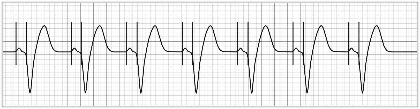
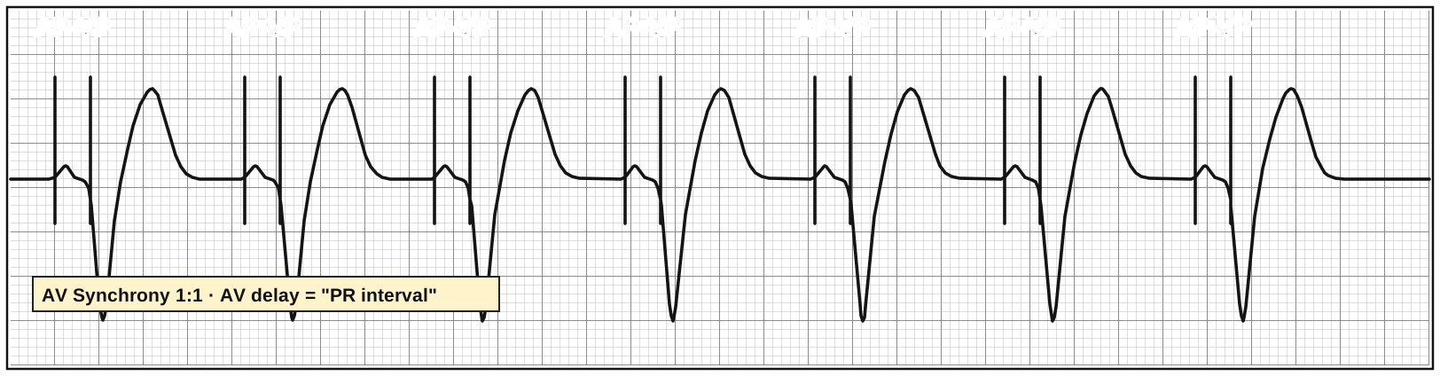
- Two spikes per beat: atrial spike → P wave, then ventricular spike → wide QRS
- With every beat paced you can't prove sensing, so this could be DDD or DOO (chamber sensed D/O, response D/O)
DDD - AS-VP → AP-VP
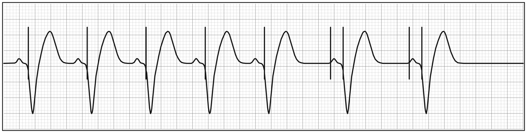
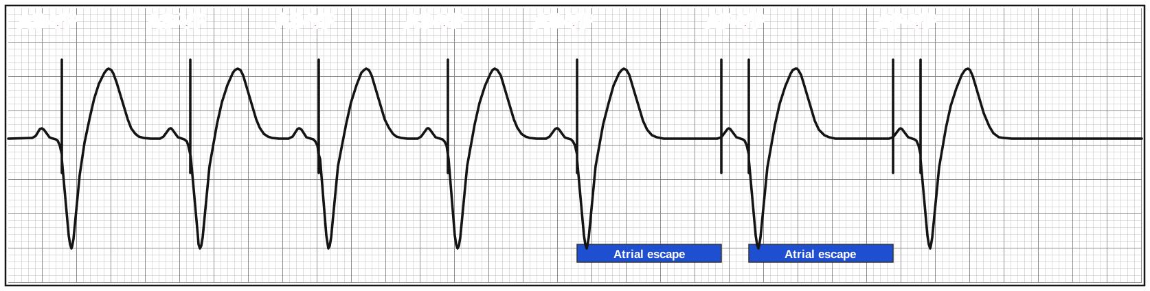
- Native P waves are sensed and tracked (AS-VP) - the device acts as an electronic AV node
- When the sinus rate slows, the atrial escape timer runs out and the device paces the atrium too (AP-VP)
- Proves the device senses and paces in both chambers → DDD
Who Gets What?
The mode follows the disease. Ask one question first: is organized atrial conduction present?
Baseline organized atrial conduction? | AV node working? | Typical patient | Mode |
No | - | Persistent/permanent AF or flutter | VVI (nothing in the atrium to sense or pace) |
Yes | Yes | Isolated sinus node dysfunction | AAI (or DDD) |
Yes | No | AV node dysfunction, paroxysmal AF | DDD |
Yes | - | Heart failure with dyssynchrony | CRT (biventricular, usually DDD-based) |
When Things Go Wrong
Failure to Capture
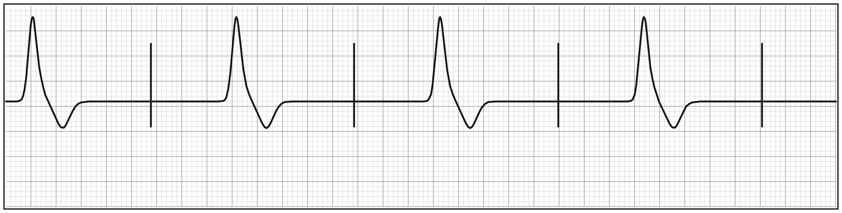
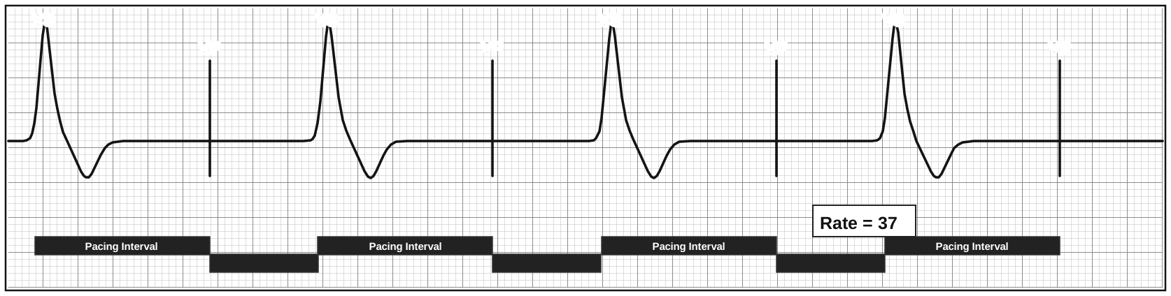
- Spike with no depolarization after it - the device fired, the heart didn't respond
- Every VP spike fails to capture; the only beats are slow ventricular escape beats (VS) that reset the timer → effective rate = 37
- If a spike falls in the refractory period right after a native beat, it can't capture - that is a sensing problem, not a capture problem
Causes of failure to capture or pace
Category | Causes |
Output failure | Output programmed too low, rising capture threshold |
Lead problems | Dislodgement, fracture, insulation breach, perforation |
Generator problem | Rare (including battery depletion) |
Why does the capture threshold rise?
- Electrolyte and metabolic problems such as hyperkalemia and acidosis
- Drugs - antiarrhythmics (especially class IC like flecainide)
- Myocardial injury - ischemia, fibrosis, infiltrative disease
- Inflammation
- Scarring at the lead tip causing exit block
Twiddler Syndrome: the patient twists the generator in its pocket (often unknowingly), coiling and retracting the leads until they dislodge or fracture. Look for coiled leads on CXR and compare lead position to a prior film.
Oversensing - T Waves
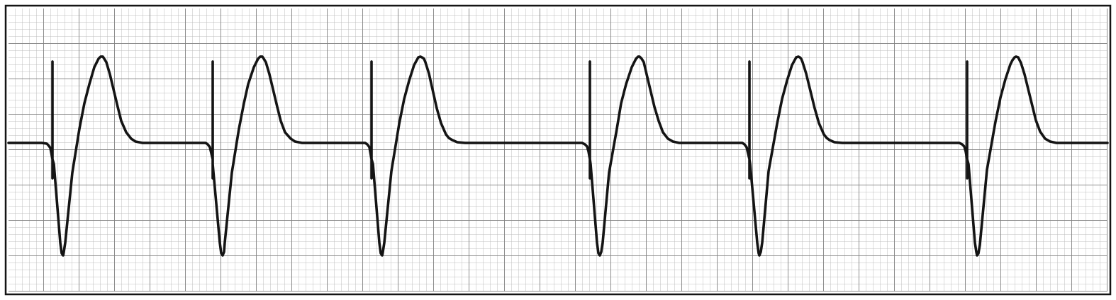
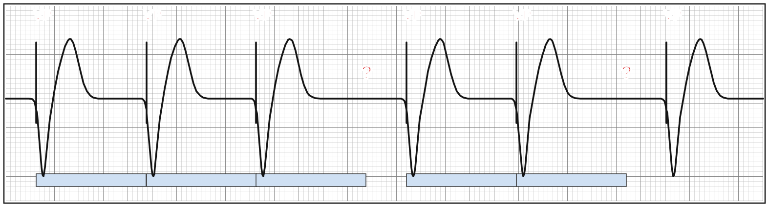
- VVI pacing with two unexplained pauses (?) - no spike where one should be
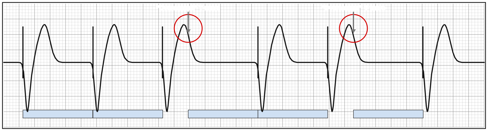
- The device is sensing its own T wave as a ventricular beat. Each "sensed" T wave resets the timer, so the next spike comes one pacing interval after the T wave
- Oversensing = the device thinks there are native beats when there are not
Oversensing - Noise
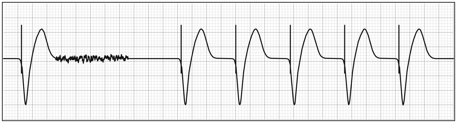
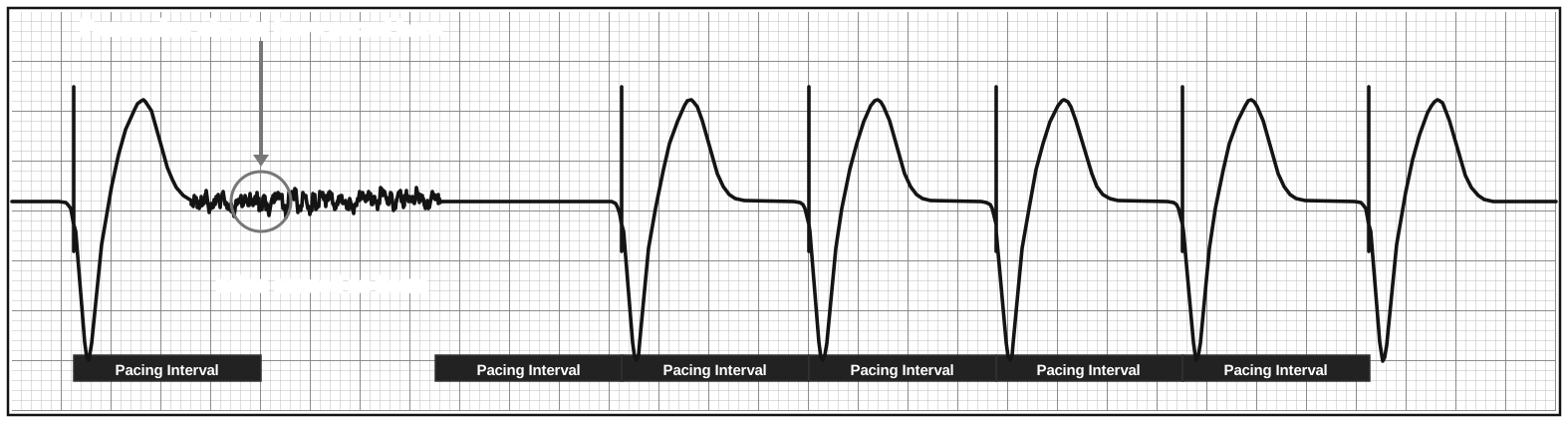
- The device saw the noisy baseline as ventricular activity and withheld pacing → pause
- Often intermittent - keep it on the differential whenever a paced patient has pauses
- Inhibited pacing → bradycardia. Triggered pacing (tracking false atrial signals) → tachycardia
Causes of oversensing
Category | Examples |
Programming too sensitive / ↑ noise | mV threshold set too low, refractory periods too short, loose or fractured lead, poor insulation |
Misreading the heart's own signals | T waves, P waves, crosstalk, double counting a wide QRS |
Extracardiac / environmental | Pectoral muscle movement, diaphragm, tremors, seizure, electrocautery |
Crosstalk
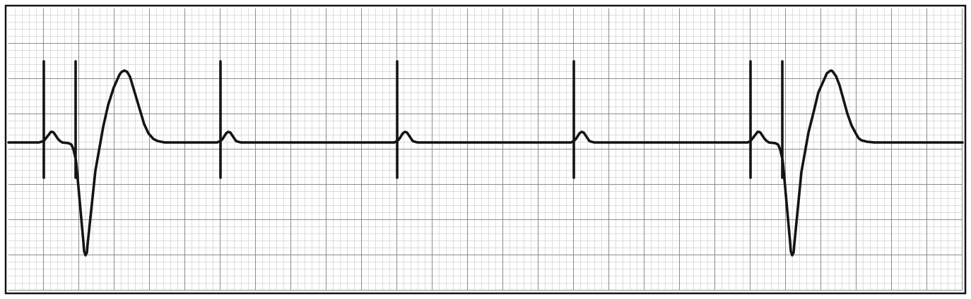
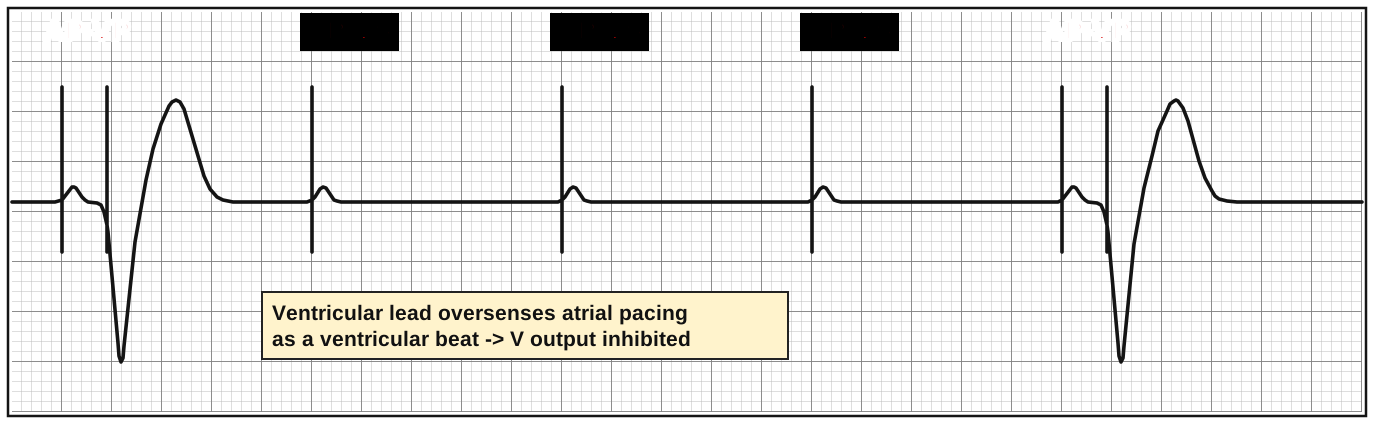
- A special type of oversensing in dual chamber devices: the ventricular lead senses the atrial pacing spike and mistakes it for a native QRS
- The device thinks each beat is AP-VS, so it withholds the ventricular spike → atrial spikes with no QRS behind them
- Dangerous in AV block - every inhibited beat is ventricular asystole
- Devices protect against it with a ventricular blanking period and ventricular safety pacing (a V spike ~100-120 ms after the A spike is safety pacing, not a malfunction)
Failure to Pace (Output Failure)
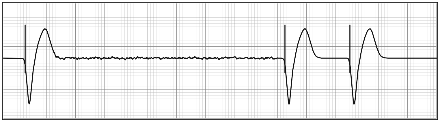
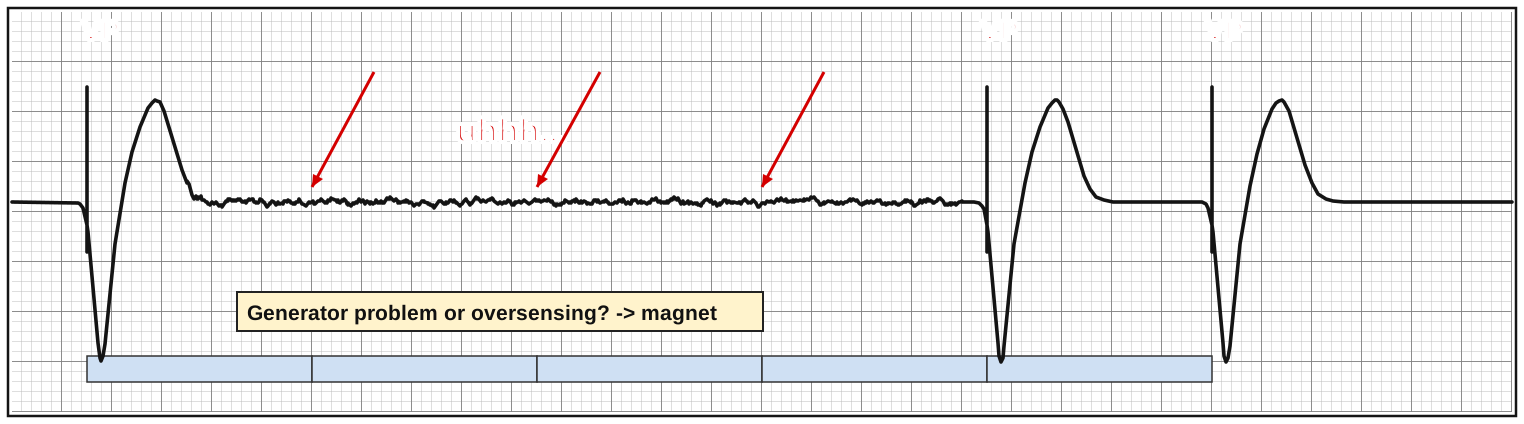
- Expected spikes never appear (red arrows) - the device didn't fire
- Causes: lead fracture, loose set screw, battery depletion, generator failure
- Is it a generator problem or an oversensing problem? Both look like a pause with no spike. Oversensing is far more common
🧲 Magnet test: a magnet turns off sensing (asynchronous pacing). If the pauses disappear → oversensing. If the pauses persist → true output failure.
Undersensing
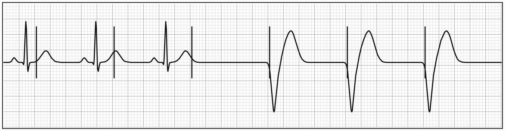
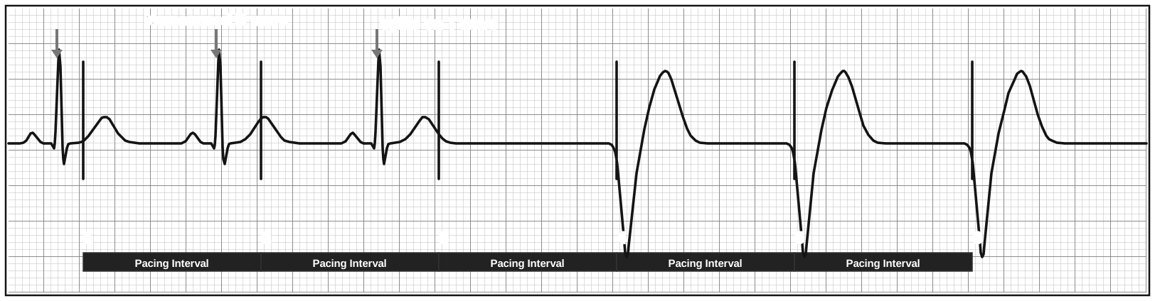
- The device can't see intrinsic depolarization - it misses true P or R waves
- It paces as if there were no underlying rhythm, behaving like an asynchronous mode (VOO, AOO, DOO)
- Look for spikes too soon after native beats, sometimes on the T wave (spike 3)
- Common causes are similar to failure to capture: smaller signals from MI, fibrosis, hyperkalemia, drugs, new BBB, lead problems, or sensitivity programmed too high
Memory aid: Oversensing → under-pacing (pauses). Undersensing → over-pacing (extra spikes). Capture and sensing problems on the same strip → think lead.
Abnormally Normal
These strips look broken but are expected device behavior.
Fusion and Pseudofusion
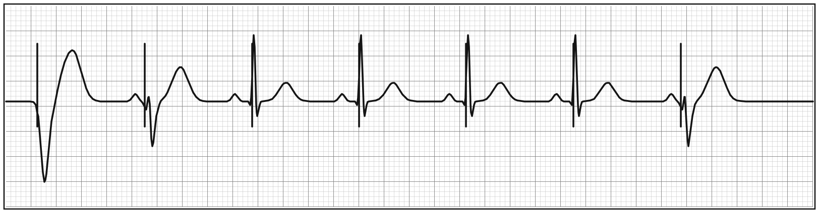
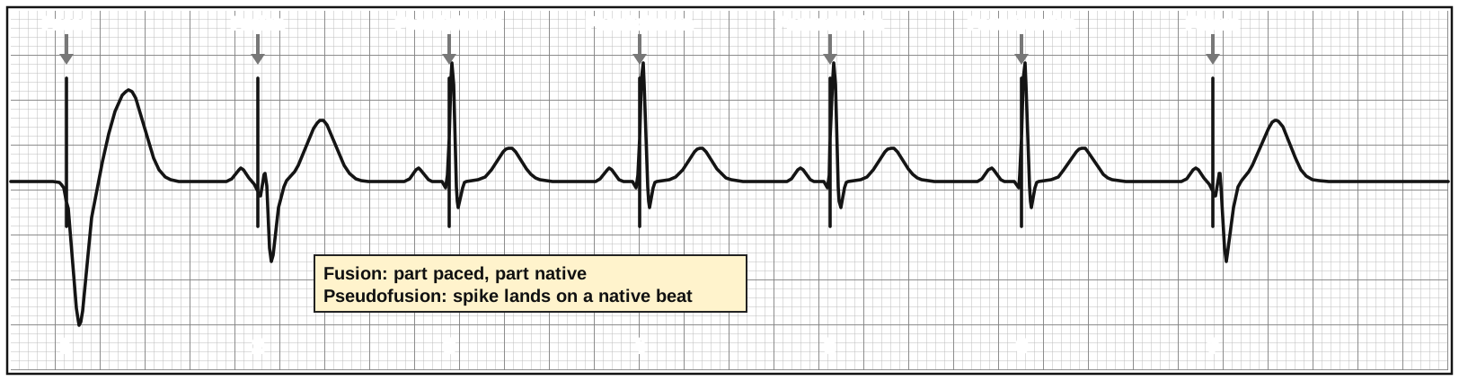
- Fusion: part paced, part native - a hybrid QRS between the two morphologies
- Pseudofusion: the spike lands on top of a native QRS that was already underway; the spike does nothing and the QRS looks native
- Happens when the native rate is close to the pacing rate. Not a malfunction
Hysteresis
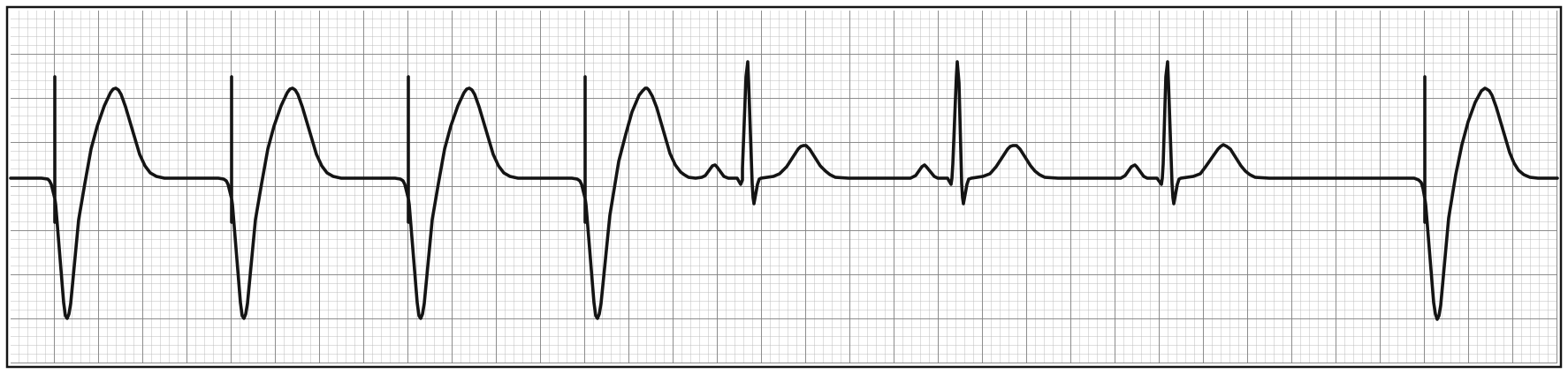
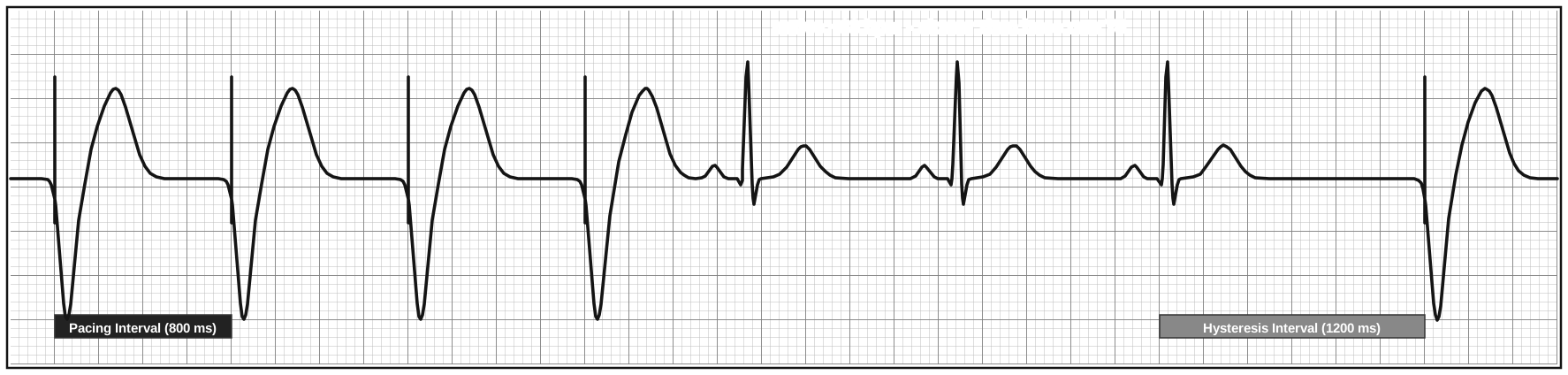
- After native beat(s), the pacemaker waits longer than its usual interval before it starts pacing
- Here the base rate is 75 (800 ms), but after native beats the device waits 1200 ms
- Gives the patient's own rhythm a chance, avoids unnecessary pacing, saves battery
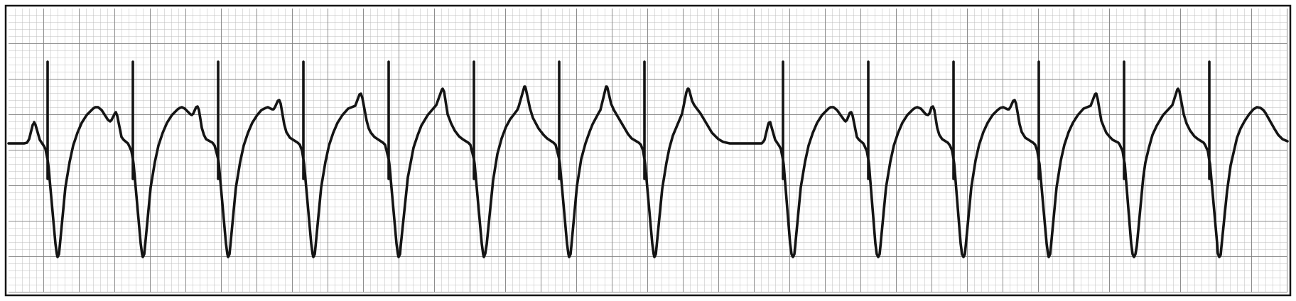
Maximum Tracking Rate (MTR)
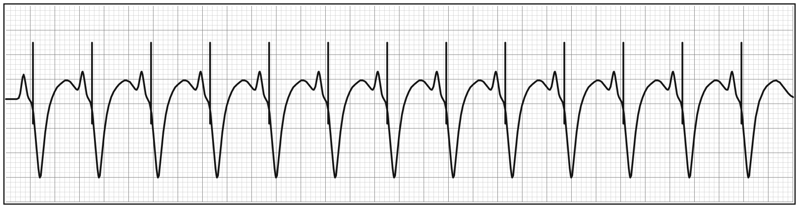
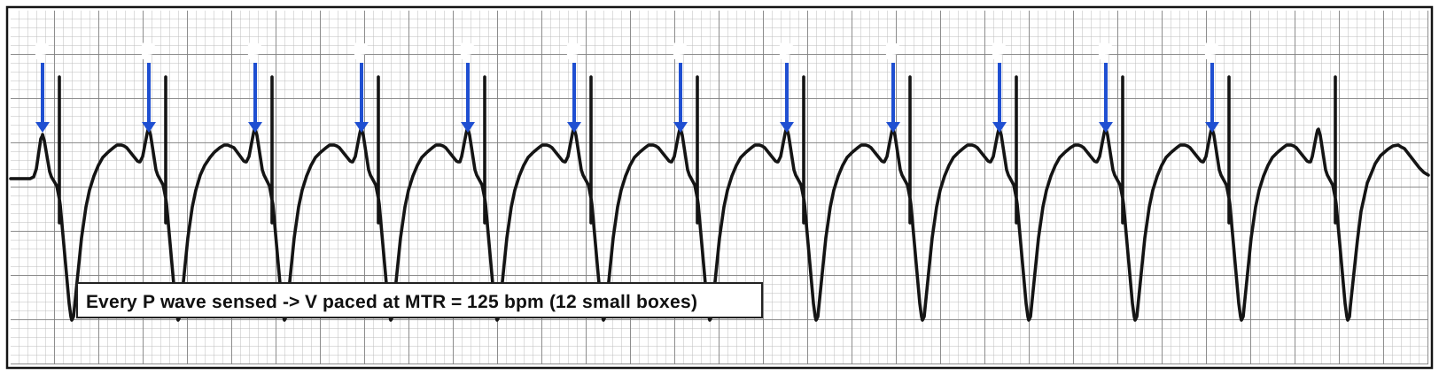
- The fastest the pacemaker will pace the ventricle in response to sensed atrial activity - a speed limit
- Every P wave (blue arrows) is sensed and tracked, but the ventricle is paced right at 125 bpm (12 small boxes) - the speed limit
PVARP and Pacemaker Wenckebach
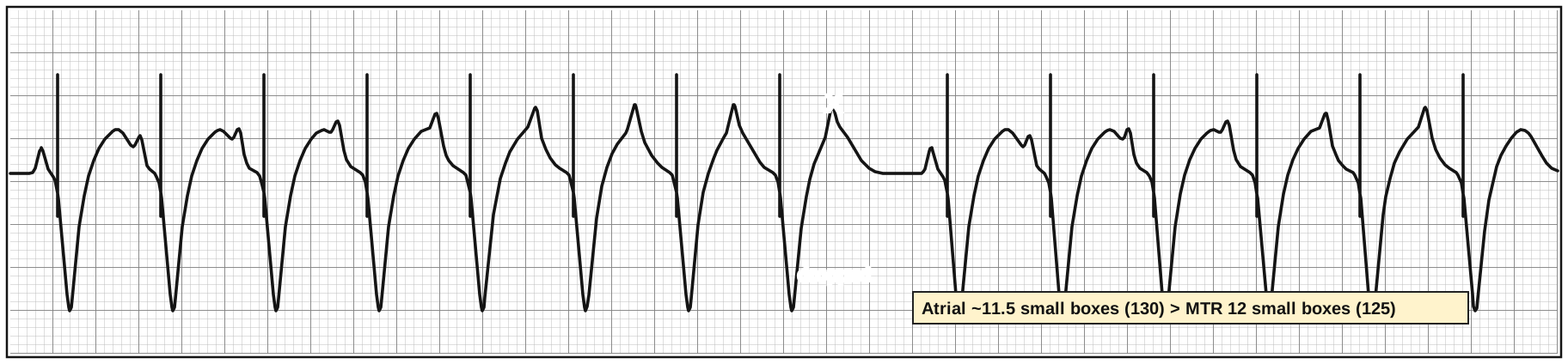
- Atrial rate (~11.5 small boxes = 130 bpm) is faster than the ventricular rate, which is stuck at the MTR (12 small boxes = 125 bpm)
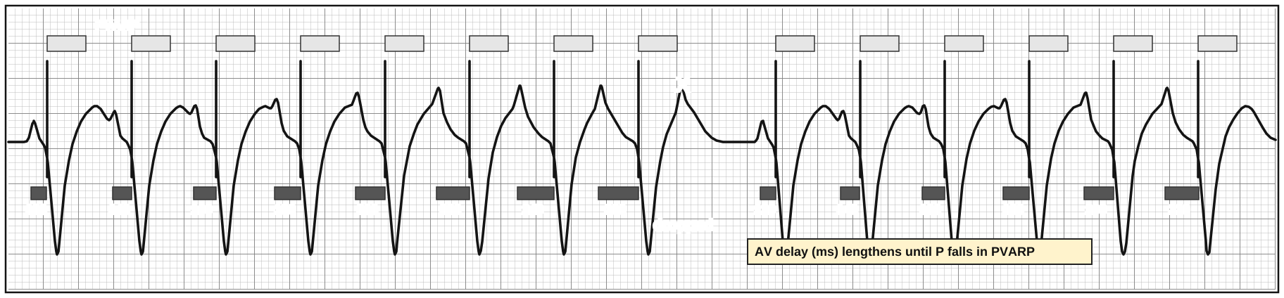
- PVARP (post-ventricular atrial refractory period): a short window after every ventricular beat where the atrial channel does not sense
- Pacemaker Wenckebach: the device prolongs the AV delay beat by beat to stay at the MTR (120 → 140 → … → 260 ms) until an atrial beat falls within PVARP (X) and is dropped - then the cycle restarts
- Group beating with progressive "PR" prolongation - looks just like Mobitz I. Not a malfunction - the real ED question is why is the atrial rate so fast?
Pacemaker Multiblock (2:1)
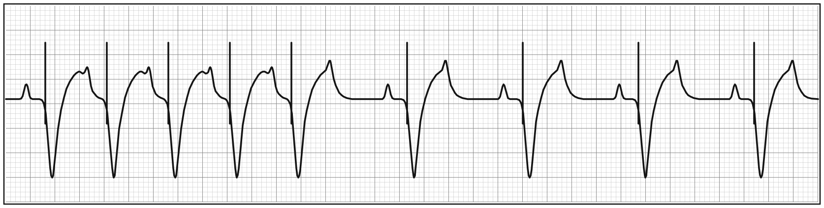
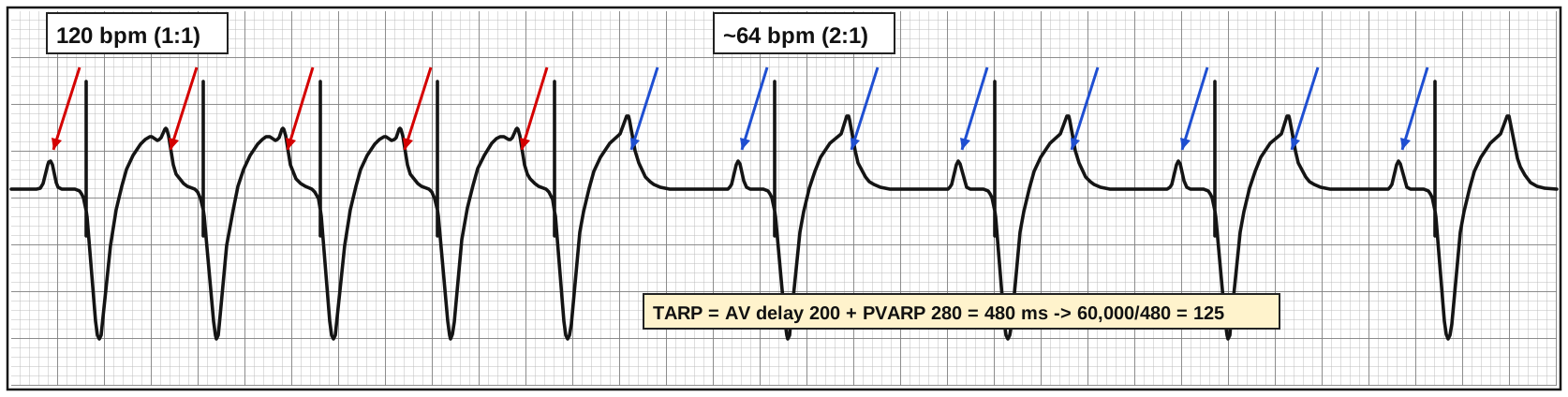
- Atrial activity is much faster than the MTR, so every other P wave falls within PVARP (blue arrows) → 2:1 conduction
- The heart rate suddenly halves (120 → 60-65) - patients feel this with exertion or fever
Calculating the atrial rate limit (2:1 point)
- TARP (total atrial refractory period) = AV delay + PVARP
- Atrial rate limit = 60,000 ÷ TARP
- Example: TARP = 200 ms + 240 ms = 440 ms → 60,000 ÷ 440 ≈ 136 bpm. Above ~136, every other P wave is ignored
Mode Switch
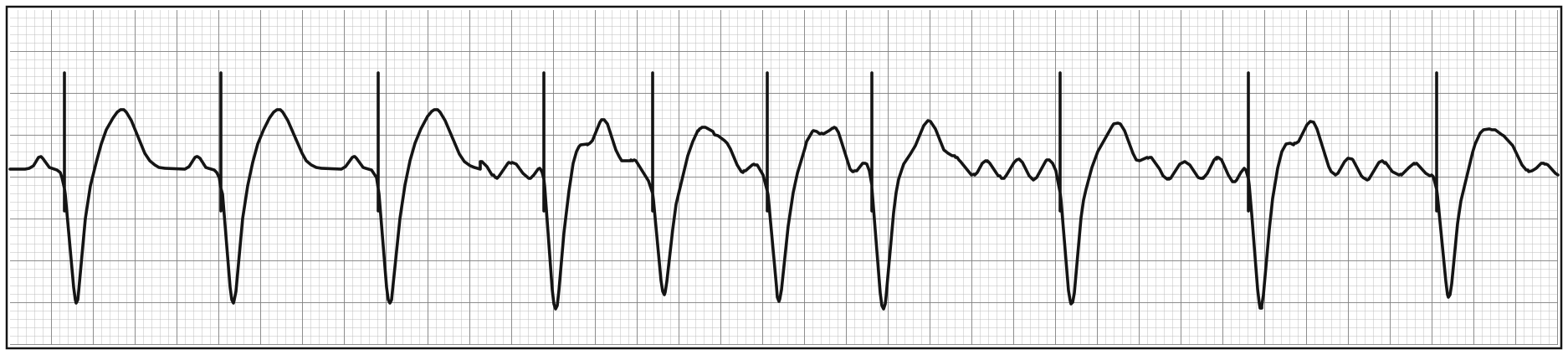
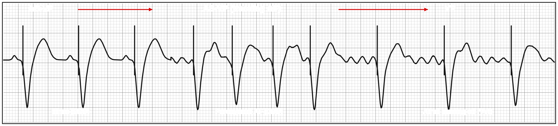
- A DDD device tracks the atrium. If the patient goes into paroxysmal AF, it briefly tracks the fibrillatory waves (fast, irregular AS-VP)
- To avoid driving the ventricle to the MTR, the device turns atrial sensing off and switches to a non-tracking mode (VVI) - regular VP at a lower rate with fibrillatory waves in the background
- When sinus rhythm returns, the device switches back to DDD
Pacemaker Mediated Tachycardia (PMT)
Triggered by Loss of Atrial Capture
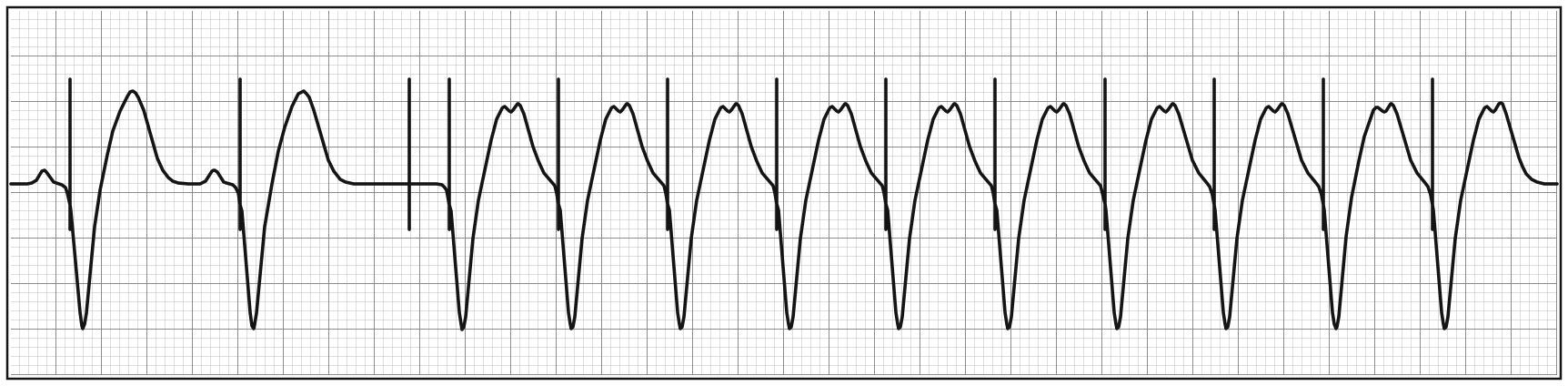
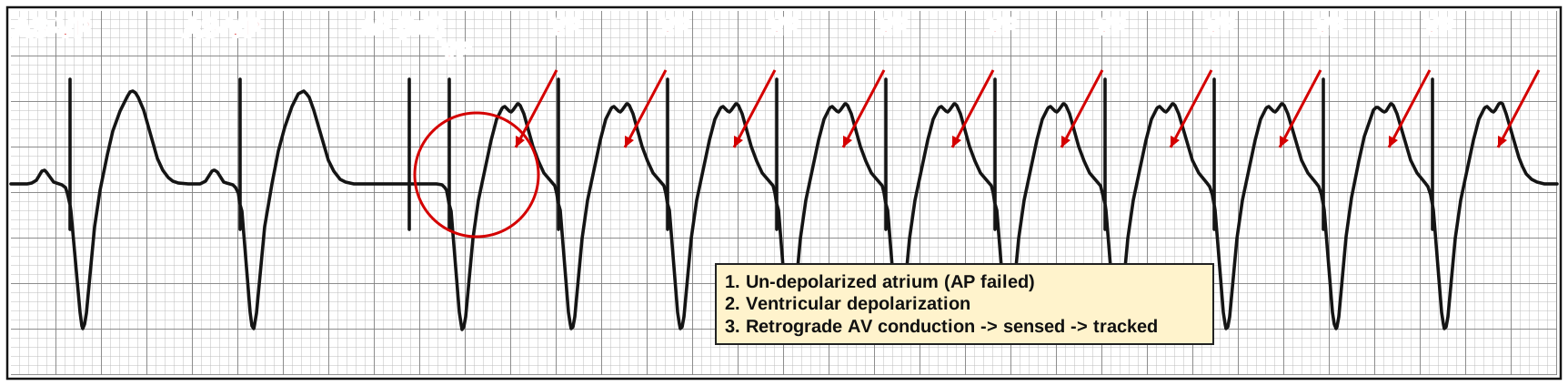
- A reentry loop where the pacemaker is part of the circuit
- The atrial spike fails to capture, so the atrium is un-depolarized when the paced ventricular beat conducts backward
- The pacemaker senses the retrograde P wave (red arrows) as a sinus P → paces the ventricle again → which conducts backward again → repeating endlessly at the maximum tracking rate
Triggered by a PVC
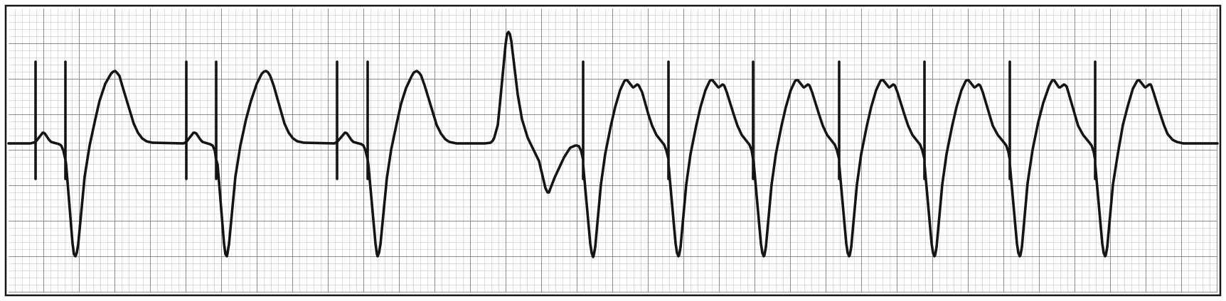
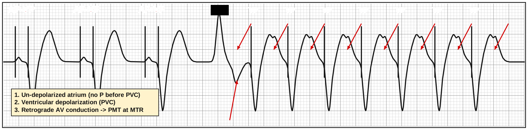
- A PVC has no preceding atrial beat, so the atrium is ready to receive a retrograde impulse - same loop
Management
- Rare - most modern devices identify and terminate PMT automatically
- Pacemaker: a magnet stops atrial sensing → the loop breaks
- ICD: a magnet does not change pacing → adenosine or vagal maneuvers to block the retrograde limb, then interrogation and reprogramming
Battery Depletion and Pacemaker Syndrome
Elective Replacement Indicator (ERI)
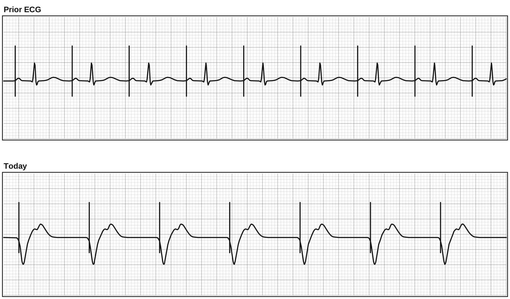
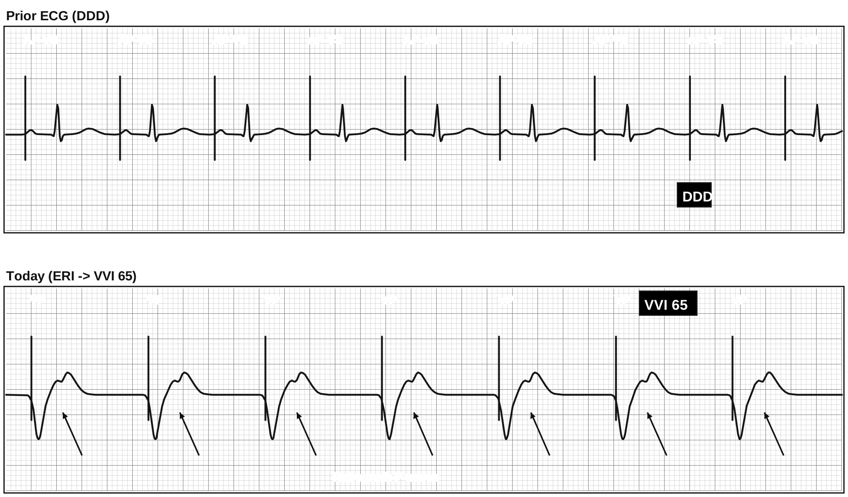
- When the battery nears the end of its life, the device reaches its elective replacement indicator (ERI) - typically about 3-6 months of battery left
- To save power, many dual chamber devices change behavior at ERI. Older Medtronic DDD devices (before ~2013), for example, switch from DDD to VVI at 65 bpm
- The prior ECG shows AP-VS (atrial paced, native QRS); today shows plain ventricular pacing with retrograde P waves - AV synchrony is lost
🔋 Why doesn't this cause PMT? Retrograde P waves are present, but atrial sensing is off in VVI. With nothing to track, the loop can't form.
Pacemaker Syndrome
Pacemaker syndrome is symptomatic AV dyssynchrony. It happens when a patient with baseline organized atrial activity is paced in VVI/VOO:
- Intact atrial conduction (DDD patient with sinus node impairment) now paced VVI
- Retrograde conduction to the atrium → atrial contraction during ventricular contraction
- It does not happen in persistent AF/flutter - there is no organized atrial contraction to lose
Mechanism → symptoms
Mechanism | Result |
Atrial contraction against closed AV valves | Venous backflow → chest discomfort, dyspnea |
Loss of atrial kick → ↓ cardiac output | Exercise intolerance, dizziness, hypotension, syncope |
Atrial distension → reflex vasodilation | Hypotension, syncope |
⚠️ ED Pearl: A paced patient with new dizziness, syncope, dyspnea, or vague chest discomfort whose ECG changed from AV synchronous pacing to plain ventricular pacing - ask when the device was last checked. Compare with an old ECG and get it interrogated.
High-Yield Pearls
- Pacemakers sense and pace. Every strip comes down to how well it does those two jobs.
- Name the mode first: chamber paced, chamber sensed, response to sense.
- Spike with no beat = failure to capture. Think output, lead, or heart (K+, drugs, ischemia).
- Pause with no spike = oversensing until proven otherwise. A magnet separates oversensing (pauses resolve) from true output failure (pauses persist).
- Spikes marching through native beats = undersensing (or a magnet/asynchronous mode).
- RV paced rhythm can hide OMI. Use the Smith-Modified Sgarbossa criteria.
- Wenckebach and 2:1 block in a DDD patient are usually expected upper-rate behavior. Find out why the atrial rate is high.
- Regular paced tachycardia at the MTR → think sinus tracking, PMT, tracked flutter/AT, or atrial oversensing. A magnet stops all of them in a pacemaker.
- New VVI-style pacing in a DDD patient + new symptoms → think battery depletion (ERI) and pacemaker syndrome.
Sources
- Kenny T. The Nuts and Bolts of Paced ECG Interpretation. Wiley-Blackwell; 2009.
- Dodd KW, Zvosec DL, Hart MA, et al. Electrocardiographic diagnosis of acute coronary occlusion myocardial infarction in ventricular paced rhythm using the modified Sgarbossa criteria. Ann Emerg Med. 2021;78(4):517-529.
- Veiga et al. 2025 (R-on-T phenomenon).
- ECG Stampede (biventricular paced morphology).
This post is for education and not medical advice.